Case Reports
. 2015 Sep 2:15:159.
doi: 10.1186/s12883-015-0415-0.

Leukoencephalopathy resolution after atypical mycobacterial treatment: a case report

Affiliations
Case Reports

Leukoencephalopathy resolution after atypical mycobacterial treatment: a case report

Marcos C B Oliveira et al. BMC Neurol. .

Abstract

Background: Association of leukoencephalopathy and atypical mycobacteriosis has been rarely reported. We present a case that is relevant for its unusual presentation and because it may shed further light on the pathogenic mechanisms underlying reversible encephalopathies.

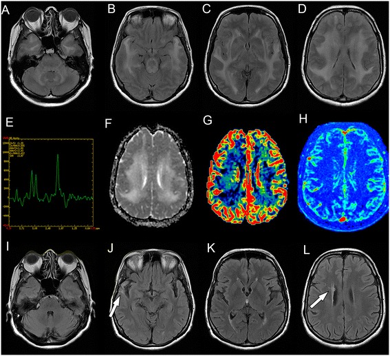
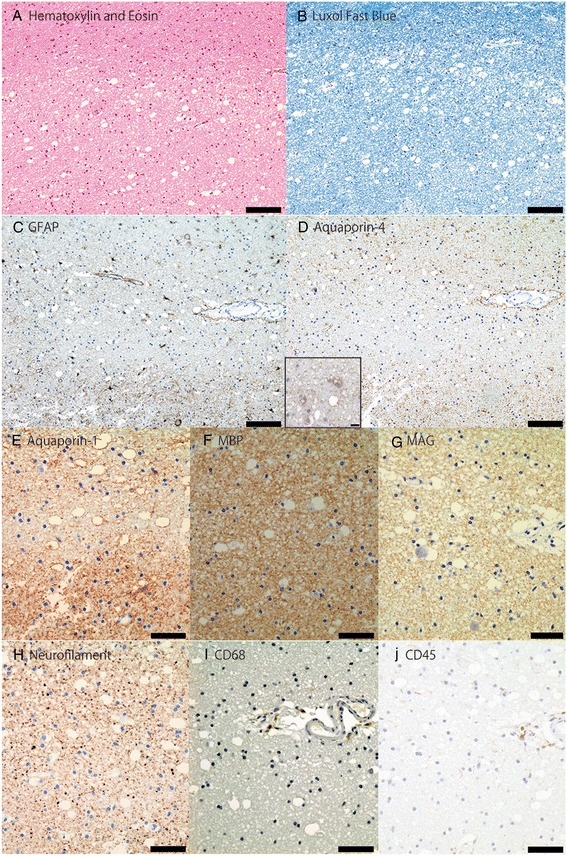
Case report: We report the case of a Hispanic 64-year-old woman with cognitive decline and extensive leukoencephalopathy. Magnetic resonance imaging revealed white-matter lesions with increased water diffusivity, without blood-brain-barrier disruption. Brain biopsy showed tissue rarefaction with vacuolation, mild inflammation, few reactive astrocytes and decreased aquaporin water-channel expression in the lesions. Six months later, she was diagnosed with atypical mycobacterial pulmonary infection. Brain lesions resolved after antimycobacterial treatment.

Conclusion: We hypothesize leukoencephalopathic changes and vasogenic edema were associated with decreased aquaporin expression. Further studies should clarify if reversible leukoencephalopathy has a causal relationship with decreased aquaporin expression and atypical mycobacterial infection, and mechanisms underlying leukoencephalopathy resolution after antimycobacterial treatment. This article may contribute to the understanding of pathogenic mechanisms underlying magnetic resonance imaging subcortical lesions and edema, which remain incompletely understood.


Figures

Fig. 1
Fig. 1
Serial brain magnetic resonance imaging studies of a patient with reversible leukoencephalopathy. Brain magnetic resonance imaging (MRI). Initial FLAIR images (a-d) show diffuse symmetrical confluent hyperintensities involving cerebral white matter, extending to the brainstem and cerebellar white matter. Note mass effect evidenced by sulci, fissure and ventricle effacement (more remarkable considering patient’s age - 64 years old). Corresponding white matter MR spectroscopy (e) (multivoxel, TE = 135 ms) demonstrates no definite metabolic changes. Apparent diffusion coefficients (ADC) map (f) demonstrates diffusion facilitation, signaling vasogenic edema. There was no contrast enhancement (not shown) or significant changes appreciated in color maps proportional to relative cerebral blood volume (rCBV) (g) obtained from a dynamic susceptibility contrast (DSC) perfusion (T2*) study. Color maps proportional to wash in rate (h) obtained from a dynamic contrast-enhanced (DCE) permeability (T1) sequence were also unremarkable. Images F-H are in the same level as D. After treatment for atypical mycobacteriosis, white matter changes disappeared, as shown in (FLAIR) images (i-l) obtained two years after the initial exam (arrows in J and L point to biopsy sites, partially characterized in these images)
Fig. 2
Fig. 2
Brain biopsy results of a patient with reversible leukoencephalopathy. a to j: Brain biopsy results of the patient with reversible leukoencephalopathy prior to atypical mycobacteria treatment shows (a) mild tissue rarefaction with vacuolation, very sparse perivascular inflammatory infiltrates, and (b) no evidence of demyelination. cd Presence of relatively few glial fibrillary acidic protein (GFAP) positive astrocytes with reduced aquaporin-4 expression in the lesion compared to the surrounding area. Scale bar = 100 μm. High magnification (400x) on D shows aquaporin-4 on the membrane of reactive astrocytes. Scale bar = 10 μm. e Aquaporin-1 expression is also reduced in the lesion, but in less extensively than aquaporin-4. fg Myelin sheath is preserved with no loss of myelin basic protein (MBP) and myelin associated glycoprotein (MAG). h No signs of neuronal or axonal damage. ij Few lymphocytes (CD45+) and macrophages (CD68+) are found in the perivascular space, while immunoglobulin and complement C9neo deposition are absent (not shown). Scale bar = 50 μm. (Magnification ad = 100x; ej = 200x)

Similar articles

Cited by

References

    1. Hinchey J, Chaves C, Appignani B, Breen J, Pao L, Wang A, et al. A reversible posterior leukoencephalopathy syndrome. N Engl J Med. 1996;334:494–500. doi: 10.1056/NEJM199602223340803. - DOI - PubMed
    1. Lee VH, Wijdicks EM, Manno EM, Rabinstein AA. Clinical spectrum of reversible posterior leukoencephalopathy syndrome. Arch Neurol. 2008;65:205–210. - PubMed
    1. Magaña SM, Matiello M, Pittock SJ, McKeon A, Lennon VA, Rabinstein AA, et al. Posterior reversible encephalopathy syndrome in neuromyelitis optica spectrum disorders. Neurology. 2009;72(8):712–717. doi: 10.1212/01.wnl.0000343001.36493.ae. - DOI - PubMed
    1. Mak A, Chan BP, Yeh IB, Ho RC, Boey ML, Feng PH, et al. Neuropsychiatric lupus and reversible posterior leucoencephalopathy syndrome: a challenging clinical dilemma. Rheumatology. 2008;47:256–262. doi: 10.1093/rheumatology/kem319. - DOI - PubMed
    1. Bartynski WS, Boardman JF, Zeigler ZR, Shadduck RK, Lister J. Posterior reversible encephalopathy syndrome in infection, sepsis, and shock. AJNR Am J Neuroradiol. 2006;27:2179–2190. - PMC - PubMed

Publication types

MeSH terms

Substances