. 2015 Sep 22;6(28):25784-800.
doi: 10.18632/oncotarget.4703.

AF4 and AF4-MLL mediate transcriptional elongation of 5-lipoxygenase mRNA by 1, 25-dihydroxyvitamin D3

Affiliations

AF4 and AF4-MLL mediate transcriptional elongation of 5-lipoxygenase mRNA by 1, 25-dihydroxyvitamin D3

Khalil Ahmad et al. Oncotarget. .

Abstract

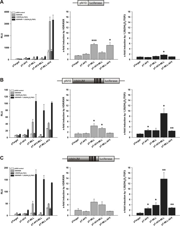
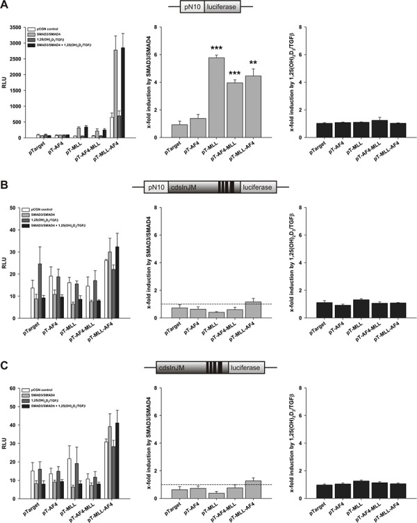
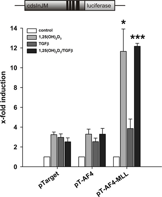
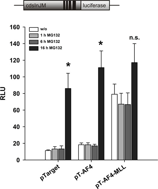
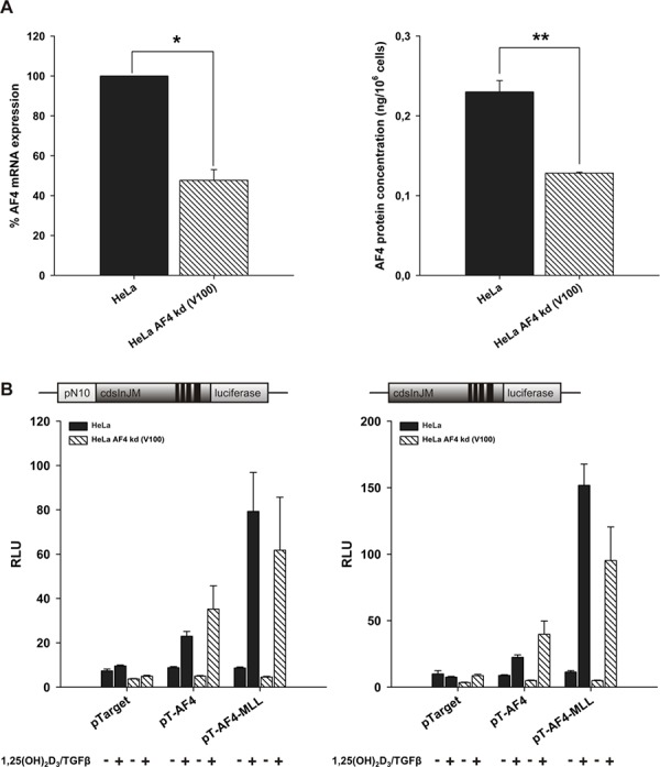
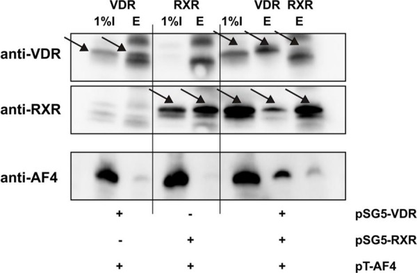
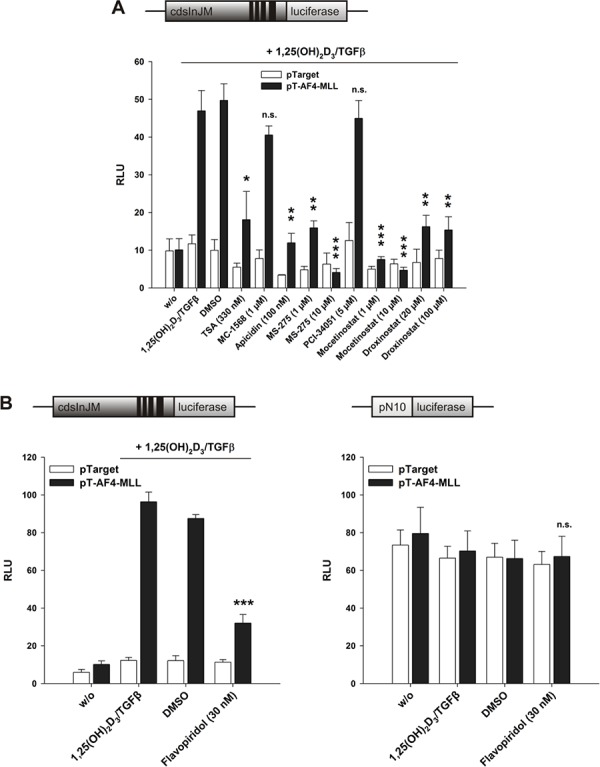
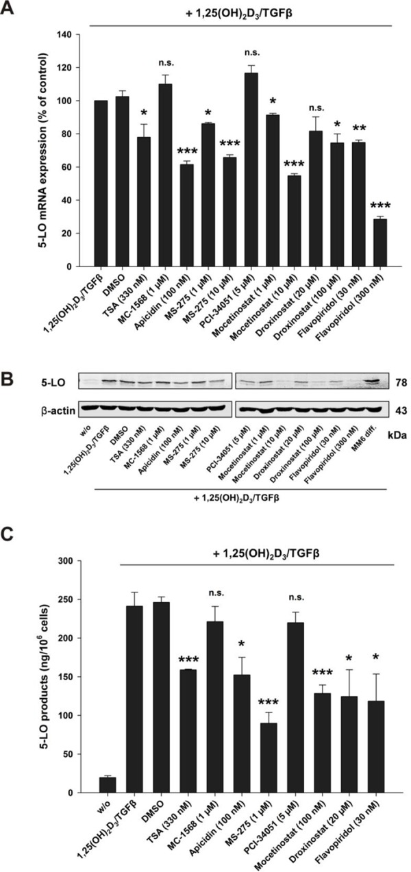
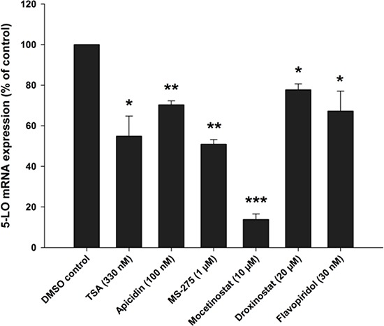
The human 5-lipoxygenase (5-LO), encoded by the ALOX5 gene, is the key enzyme in the formation of pro-inflammatory leukotrienes. ALOX5 gene transcription is strongly stimulated by calcitriol (1α, 25-dihydroxyvitamin D3) and TGFβ (transforming growth factor-β). Here, we investigated the influence of MLL (activator of transcript initiation), AF4 (activator of transcriptional elongation) as well as of the leukemogenic fusion proteins MLL-AF4 (ectopic activator of transcript initiation) and AF4-MLL (ectopic activator of transcriptional elongation) on calcitriol/TGFβ-dependent 5-LO transcript elongation. We present evidence that the AF4 complex directly interacts with the vitamin D receptor (VDR) and promotes calcitriol-dependent ALOX5 transcript elongation. Activation of transcript elongation was strongly enhanced by the AF4-MLL fusion protein but was sensitive to Flavopiridol. By contrast, MLL-AF4 displayed no effect on transcriptional elongation. Furthermore, HDAC class I inhibitors inhibited the ectopic effects caused by AF4-MLL on transcriptional elongation, suggesting that HDAC class I inhibitors are potential therapeutics for the treatment of t(4;11)(q21;q23) leukemia.

Keywords: 5-lipoxygenase; AF4; HDAC; MLL; calcitriol.


Conflict of interest statement

CONFLICT OF INTEREST

The authors declare no conflict of interest.

Figures

Figure 1
Figure 1. Schematic representation of applied reporter gene constructs in the reporter gene assay experiments
Figure 2
Figure 2. Effect of AF4, MLL, AF4-MLL and MLL-AF4 overexpression on VDR/RXR- and calcitriol/TGFβ-dependent response of the reporter gene constructs A. pN10 B. pN10cdsInJM and C. pGL3cdsInJM
In order to study inducibility of the constructs by calcitriol/TGFβ, HeLa cells were co-transfected either with the pSG5 control vector or with the expression vectors for the nuclear receptors VDR and RXR. 16 hours after transfection, cells were incubated without or with calcitriol (50 nM) and TGFβ (1 ng/ml). After 40 hours, luciferase activity was determined. Results are given as RLU (left panels) which include normalization to transfection efficiency and as x-fold induction by VDR/RXR over pSG5 control (middle panels) or x-fold induction by calcitriol/TGFβ over VDR/RXR (right panels). Each experiment was performed in triplicate. Results are presented as mean + S.E.M. of three independent experiments. Two-tailed t-test was performed to determine significant differences in relation to co-transfection of the backbone vector pTarget, indicated by asterisks (*P < 0.05, **P < 0.01, ***P < 0.001).
Figure 3
Figure 3. Effect of AF4, MLL, AF4-MLL and MLL-AF4 overexpression on SMAD3/SMAD4- and calcitriol/TGFβ-dependent response of the reporter gene constructs A. pN10 B. pN10cdsInJM and C. pGL3cdsInJM
In order to study inducibility of the constructs by calcitriol/TGFβ, HeLa cells were co-transfected either with the pCGN control vector or with the expression vectors for the nuclear receptors SMAD3 and SMAD4. 16 hours after transfection, cells were incubated without or with calcitriol (50 nM) and TGFβ (1 ng/ml). After 40 hours, luciferase activity was determined. Results are given as RLU (left panels) which include normalization to transfection efficiency and as x-fold induction by SMAD3/SMAD4 over pCGN control (middle panels) or x-fold induction by calcitriol/TGFβ over SMAD3/SMAD4 (right panels). Each experiment was performed in triplicate. Results are presented as mean − S.E.M. of three independent experiments. Two-tailed t-test was performed to determine significant differences in relation to co-transfection of the backbone vector pTarget, indicated by asterisks (*P < 0.05, **P < 0.01, ***P < 0.001).
Figure 4
Figure 4. Contribution of calcitriol and TGFβ to the AF4-MLL-dependent reporter gene activation by calcitriol/TGFβ
HeLa cells were transfected with the pGL3cdsInJM reporter gene construct as well as the expression vectors for the nuclear receptors VDR and RXR and were co-transfected either with the pTarget control vector or the expression vectors for AF4 or AF4-MLL as indicated. 16 hours after transfection, cells were incubated without or with calcitriol (50 nM) and/or TGFβ (1 ng/ml). After 40 hours, luciferase activity was determined and results are given as x-fold induction over untreated cells. Each experiment was performed in triplicate. Results are presented as mean − S.E.M. of three independent experiments. Two-tailed t-test was performed to determine significant differences in relation to co-transfection of the backbone vector pTarget, indicated by asterisks (*P < 0.05, **P < 0.01, ***P < 0.001).
Figure 5
Figure 5. Effect of the proteasome inhibitor MG132 on reporter gene activation by AF4 and AF4-MLL
HeLa cells were transfected with the pGL3cdsInJM reporter gene construct and the expression vectors for the nuclear receptors VDR and RXR and were co-transfected either with the pTarget control vector or the expression vectors for AF4 or AF4-MLL. 16 hours after transfection, cells were incubated with calcitriol (50 nM) and TGFβ (1 ng/ml). Cells were treated with MG132 (5 μM) for 1, 6 and 16 hours. 40 hours after transfection, luciferase activity was determined and the results are given as RLU which include normalization to transfection efficiency. Each experiment was performed in triplicate. Results are presented as mean − S.E.M. of three independent experiments. Two-tailed t-test was performed to determine the significance in relation to MG132 untreated cells, indicated by asterisks (*P < 0.05, **P < 0.01, ***P < 0.001).
Figure 6
Figure 6. Effect of stable AF4 knockdown in HeLa cells
A. AF4 knockdown cells were generated with lentiviral particles, produced by co-transfection of HEK293T cells with the shRNA plasmid for AF4, pCMV-dR8.91 and pMD2.G. Knockdown efficiency of AF4 was checked by qPCR, using β-actin as housekeeping gene, and viaAFF1 ELISA Kit in three independent experiments, respectively. One sample t-test was performed to determine the significance of AF4 knockdown on mRNA level (left graph) and two-tailed t-test on protein level (right graph) (*P < 0.05, **P < 0.01, ***P < 0.001). B. HeLa wild type and HeLa AF4 knockdown cells were co-transfected with the pN10cdsInJM or pGL3cdsInJM reporter gene construct, pTarget or the expression vectors for AF4 or AF4-MLL and with the expression vectors for the nuclear receptors VDR and RXR. 16 hours after transfection, cells were incubated without or with calcitriol (50 nM) and TGFβ (1 ng/ml) as indicated. After 40 hours, luciferase activity was determined and is given as RLU which include normalization to transfection efficiency. Each experiment was performed in triplicate. Results are presented as mean + S.E.M. of three independent experiments.
Figure 7
Figure 7. Co-immunoprecipitation of AF4 and VDR/RXR
HEK293T cells were co-transfected with the expression vectors for AF4 and the nuclear receptors VDR and/or RXR as indicated. Then, cells were cultured for 48 hours. After harvest and cell lysis, immunoprecipitation was performed with VDR or RXR antibodies as indicated. Subsequently, supernatants (I) and bead eluates (E) were analyzed by Western blot analysis for VDR, RXR and AF4. Figure illustrates one representative Co-IP of three independent experiments.
Figure 8
Figure 8. Effect of A. HDAC inhibitors and B. CDK9 inhibitor Flavopiridol on the AF4-MLL-dependent induction of reporter gene activity by calcitriol/TGFβ
HeLa cells were transfected with the pGL3cdsInJM reporter gene construct plus the expression vectors for VDR and RXR (left panel) or the pN10 reporter gene construct (right panel). Additionally, cells were co-transfected either with the pTarget control vector or the expression vector for AF4-MLL as indicated. 16 hours after transfection, cells were incubated without or with calcitriol (50 nM) and TGFβ (1 ng/ml) and the indicated HDAC inhibitors or the CDK9 inhibitor Flavopiridol. After 40 hours, luciferase activity was determined. Results are given as RLU which include normalization to transfection efficiency. Each experiment was performed in triplicate. Results are presented as mean + S.E.M. of three independent experiments. Two-tailed t-test was performed to determine the significance of inhibitor treatment, indicated by asterisks (*P < 0.05, **P < 0.01, ***P < 0.001).
Figure 9
Figure 9. Inhibition of A. 5-LO mRNA expression, B. protein expression and C. activity by class I HDAC inhibitors or the CDK9 inhibitor Flavopiridol in MM6 cells
MM6 cells were incubated without or with calcitriol (50 nM) and TGFβ (1 ng/ml) and in the presence of HDAC inhibitors or the CDK9 inhibitor Flavopiridol as indicated. After 24 hours, cells were harvested and 5-LO mRNA and protein expression were determined by real-time PCR and by Western blot analysis, respectively. The MM6 diff sample in panel B is derived from cells incubated for 96 hours with calcitriol/TGFβ. Cellular 5-LO activity was determined after cell differentiation by calcitriol/TGFβ in the presence of the indicated HDAC inhibitors for 96 hours. In each case three independent experiments were performed. Two-tailed t-test was performed to determine the significance of inhibitor treatment, indicated by asterisks (*P < 0.05, **P < 0.01, ***P < 0.001).
Figure 10
Figure 10. Inhibition of 5-LO mRNA expression by class I HDAC inhibitors or the CDK9 inhibitor Flavopiridol in MV4-11 cells
MV4-11 cells were differentiated with (50 nM) and TGFβ (1 ng/ml) and cultured in the presence of HDAC inhibitors or the CDK9 inhibitor Flavopiridol as indicated. After 24 hours, cells were harvested and 5-LO mRNA expression was determined by real-time PCR in three independent experiments. One sample t-test was performed to determine the significance of inhibitor treatment, indicated by asterisks (*P < 0.05, **P < 0.01, ***P < 0.001).

References

    1. Rådmark O, Werz O, Steinhilber D, Samuelsson B. 5-Lipoxygenase: regulation of expression and enzyme activity. Trends Biochem Sci. 2007;32:332–341. - PubMed
    1. Funk CD, Hoshiko S, Matsumoto T, Rådmark O, Samuelsson B. Characterization of the human 5-lipoxygenase gene. Proc Natl Acad Sci U S A. 1989;86:2587–2591. - PMC - PubMed
    1. Hoshiko S, Rådmark O, Samuelsson B. Characterization of the human 5-lipoxygenase gene promoter. Proc Natl Acad Sci U S A. 1990;87:9073–9077. - PMC - PubMed
    1. Klan N, Seuter S, Schnur N, Jung M, Steinhilber D. Trichostatin A and structurally related histone deacetylase inhibitors induce 5-lipoxygenase promoter activity. Biol Chem. 2003;384:777–785. - PubMed
    1. Ahmad K, Katryniok C, Scholz B, Merkens J, Löscher D, Marschalek R, Steinhilber D. Inhibition of class I HDACs abrogates the dominant effect of MLL-AF4 by activation of wild-type MLL. Oncogenesis. 2014;3:e127. - PMC - PubMed

Publication types

MeSH terms

Substances