. 2015 Sep 2:5:13608.
doi: 10.1038/srep13608.

Evidence for the expression of abundant microRNAs in the locust genome

Affiliations

Evidence for the expression of abundant microRNAs in the locust genome

Yanli Wang et al. Sci Rep. .

Abstract

Substantial accumulation of neutral sequences accounts for genome size expansion in animal genomes. Numerous novel microRNAs (miRNAs), which evolve in a birth and death manner, are considered evolutionary neutral sequences. The migratory locust is an ideal model to determine whether large genomes contain abundant neutral miRNAs because of its large genome size. A total of 833 miRNAs were discovered, and several miRNAs were randomly chosen for validation by Northern blot and RIP-qPCR. Three additional verification methods, namely, processing-dependent methods of miRNA biogenesis using RNAi, evolutionary comparison with closely related species, and evidence supported by tissue-specific expression, were applied to provide compelling results that support the authenticity of locust miRNAs. We observed that abundant local duplication events of miRNAs, which were unique in locusts compared with those in other insects with small genome sizes, may be responsible for the substantial acquisition of miRNAs in locusts. Together, multiple evidence showed that the locust genome experienced a burst of miRNA acquisition, suggesting that genome size expansion may have considerable influences of miRNA innovation. These results provide new insight into the genomic dynamics of miRNA repertoires under genome size evolution.


Figures

Figure 1
Figure 1. Number of miRNA precursors in the locust genome.
(A) Summary of identified miRNA precursors in the present study and previous study. (B) Summary of identified miRNA precursors from miRBase in metazoan species. The vertebrate and insect miRNA precursors are the predominant representatives (~84%) of metazoan miRNA precursors in miRBase Release 21. The numbers of miRNA precursors at the top ranks are shown in insects and vertebrates.
Figure 2
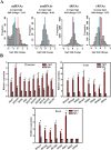
Figure 2. MiRNA validation using processing-dependent methods of miRNA biogenesis.
(A) Effects of Drosha knockdown using RNAi on miRNA biogenesis. MiRNA expression was determined with small RNA sequencing for silenced and negative control tissues. The density distribution of log2 fold changes in the expression for each miRNA between silenced and negative control tissues is shown. (B) RIP was performed with an anti-Ago1 antibody, and IgG was used as a negative control. QPCR analysis was performed to amplify miRNAs from the Ago-1 immunoprecipitates from extracts of pronotums, testes and brains. The data for the RIP assay are presented as the mean 6 SEM (n = 6). *indicates P < 0.05.
Figure 3
Figure 3. Evidence for the presence of numerous locust miRNAs in an evolutionary view.
(A) Box plot of miRNA expression for evolutionarily conserved miRNAs and lineage-specific miRNAs. *indicates P < 0.001 (Mann–Whitney–Wilcoxon tests). (B) Box plot of lineage-specific miRNA expression for lineage-specific miRNAs that showed no variations and those with sequence variations. The variable group represents the lineage-specific miRNAs in locusts that differed from those in band-winged grasshoppers. *indicates P = 0.009 (Mann–Whitney–Wilcoxon tests). (C) Effects of Drosha knockdown using RNAi for miRNAs of the three different categories, namely evolutionarily conserved miRNAs, lineage-specific miRNAs with moderate/high expression and lineage-specific miRNAs with low expression. (D) Inferred MFEs of the lineage-specific miRNAs were similar to those of the evolutionarily conserved miRNAs, and significantly stronger than the binding of shuffled control sequences. (E,F) The miRNA 5′ end and 3′ end processing precision of evolutionarily conserved and lineage-specific miRNAs. The processing precision was calculated as the fraction of mapped reads that corresponded precisely to the consensus sequences of genomic locus. The miRNAs are shown on the x-axis and ordered by the processing precision. The miRNA with the most precision is at percentile 1, and the one with the least precision is at percentile 100.
Figure 4
Figure 4. Abundant tissue-specific expression of locust lineage-specific miRNAs.
(A) ID1685-5P is an example of tissue-specific miRNA, because it shows significantly higher expression in the testes compared with those in other tissues. (B) Numerous miRNAs that showed strong specificity in multiply tissues were determined by comparing the expression between the candidate tissue and other condition-specific tissues/developmental stages using statistical differential expression analysis of the edgeR program. (C) The majority of miRNAs that showed tissue-specific expression were lineage-specific miRNAs. (D) The predominant portion of locust lineage-specific miRNAs showed a tissue-specific manner in the testes or brains.
Figure 5
Figure 5. Numerous miRNAs arise from local duplication events.
(A) A large fraction of miRNA precursors showed sequence similarity to each other. (B) Six miRNA precursors that showed sequence similarity were located close to each other in a neighboring region, which suggested that they emerged from local duplication events. (C) These six miRNA precursors comprised members of different seed families, although they showed sequence similarity to each other. (D) Variations in the six miRNA precursors resulted in fluctuations in the MFEs, thereby influencing the stability of the hairpin structures of miRNA precursors. (E) The global–local alignment searches, in which all locust miRNA precursors (termed as original miRNAs) were used, were performed to identify sequences of similarity in their genomic flanking regions (duplication events). The distances between similar copies and original miRNAs were calculated for each miRNA, and the hits for the original miRNA itself were excluded in distance calculation. L. migratoria, Locusta migratoria; B. mori, Bombyx mori; A. pisum, Acyrthosiphon pisum; T. castaneum, Tribolium castaneum; D. melanogaster, Drosophila melanogaster; A. mellifera, Apis mellifera; A. aegypti, Aedes aegypti; A. gambiae, Anopheles gambiae.

Similar articles

Cited by

References

    1. Carthew R. W. & Sontheimer E. J. Origins and Mechanisms of miRNAs and siRNAs. Cell 136, 642–655, 10.1016/j.cell.2009.01.035 (2009). - DOI - PMC - PubMed
    1. Pasquinelli A. E. MicroRNAs and their targets: recognition, regulation and an emerging reciprocal relationship. Nat Rev Genet 13, 271–282, 10.1038/nrg3162 (2012). - DOI - PubMed
    1. Ameres S. L. & Zamore P. D. Diversifying microRNA sequence and function. Nat Rev Mol Cell Biol 14, 475–488, 10.1038/nrm3611 (2013). - DOI - PubMed
    1. Chekulaeva M. & Filipowicz W. Mechanisms of miRNA-mediated post-transcriptional regulation in animal cells. Current opinion in cell biology 21, 452–460, 10.1016/j.ceb.2009.04.009 (2009). - DOI - PubMed
    1. Forman J. J., Legesse-Miller A. & Coller H. A. A search for conserved sequences in coding regions reveals that the let-7 microRNA targets Dicer within its coding sequence. Proc Natl Acad Sci USA 105, 14879–14884, 10.1073/pnas.0803230105 (2008). - DOI - PMC - PubMed

Publication types

LinkOut - more resources