. 2015 Sep 1;75(17):3529-42.
doi: 10.1158/0008-5472.CAN-14-2645.

Pancreatic Cancer Cell Migration and Metastasis Is Regulated by Chemokine-Biased Agonism and Bioenergetic Signaling

Affiliations

Pancreatic Cancer Cell Migration and Metastasis Is Regulated by Chemokine-Biased Agonism and Bioenergetic Signaling

Ishan Roy et al. Cancer Res. .

Abstract

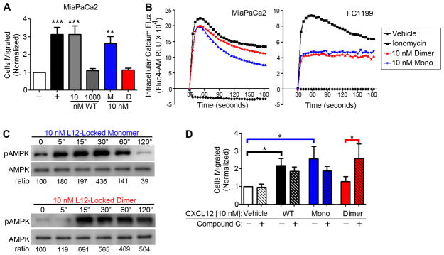
Patients with pancreatic ductal adenocarcinoma (PDAC) invariably succumb to metastatic disease, but the underlying mechanisms that regulate PDAC cell movement and metastasis remain little understood. In this study, we investigated the effects of the chemokine gene CXCL12, which is silenced in PDAC tumors, yet is sufficient to suppress growth and metastasis when re-expressed. Chemokines like CXCL12 regulate cell movement in a biphasic pattern, with peak migration typically in the low nanomolar concentration range. Herein, we tested the hypothesis that the biphasic cell migration pattern induced by CXCL12 reflected a biased agonist bioenergetic signaling that might be exploited to interfere with PDAC metastasis. In human and murine PDAC cell models, we observed that nonmigratory doses of CXCL12 were sufficient to decrease oxidative phosphorylation and glycolytic capacity and to increase levels of phosphorylated forms of the master metabolic kinase AMPK. Those same doses of CXCL12 locked myosin light chain into a phosphorylated state, thereby decreasing F-actin polymerization and preventing cell migration in a manner dependent upon AMPK and the calcium-dependent kinase CAMKII. Notably, at elevated concentrations of CXCL12 that were insufficient to trigger chemotaxis of PDAC cells, AMPK blockade resulted in increased cell movement. In two preclinical mouse models of PDAC, administration of CXCL12 decreased tumor dissemination, supporting our hypothesis that chemokine-biased agonist signaling may offer a useful therapeutic strategy. Our results offer a mechanistic rationale for further investigation of CXCL12 as a potential therapy to prevent or treat PDAC metastasis.


Conflict of interest statement

Conflicts of Interest: Michael Dwinell and Brian Volkman are founders of Protein Foundry, LLC which produces ultra-pure recombinant chemokines for research (proteinfoundry.com).

Figures

Figure 1
Figure 1. Systemic treatment with recombinant CXCL12 blocks pancreatic cancer metastasis in orthotopic xenograft mouse models
(A) Panc1 and MiaPaCa2 firefly luciferase transfected cells were orthotopically implanted into the pancreata of immunocompromised mice. At day 7 post-implantation, mice were sorted into vehicle or 5 μM CXCL12 treatment groups and were injected intraperitoneally twice-weekly and thereafter until study end. Tumor growth and dissemination were tracked weekly using bioluminescence imaging. The MiaPaCa2 model was ended at 70 days while the Panc1 model was performed as a survival study with animal removal at signs of morbidity. (B) Representative mice with Panc1 implants are shown, with CXCL12 treated mice exhibiting less tumor dissemination at day 112. (C) Tumor dissemination and metastasis was analyzed ex vivo and showed that, in both MiaPaCa2 and Panc1 models, CXCL12 treatment significantly decreased pancreatic cancer cell metastasis. (D) Representative bioluminescence image and heat-map of excised livers from Panc1 engrafted vehicle and CXCL12-treated groups, reflecting extent of metastasis. (*) denotes P≤0.05. Values = mean ± SE. n=5–6.
Figure 2
Figure 2. Ataxic doses of CXCL12 alter pancreatic cancer cell bioenergy levels and stimulate prolonged AMP-Kinase activity
(A–C) MiaPaCa2 cells were serum-starved and treated with a concentration curve ranging from chemotactic (10 nM) to ataxic (1000 nM) doses of CXCL12. Upon treatment, basal oxygen consumption and extracellular acidification rates (OCR & ECAR) were measured using a Seahorse-XF analyzer continuously for 1 hour. At 1 hour, energetic stress tests were employed sequentially, with oligomycin first used to measure ATP-linked OCR or ECAR reserve, then dinitrophenol used second to measure OCR Reserve. (*) denotes P≤0.05, (**) denotes P≤0.01. Values = mean ± SE, n=5. (D–E) Human MiaPaCa2 or patient-derived MCW512 (MCW-4), or murine KRasLSL.G12D/+-p53R172H/+-PdxCretg/+ (KPC) derived FC1199 cells were stimulated for either 15 minutes or 1 hour with 1, 10, 100, or 1000 nM recombinant CXCL12. At each time point, protein lysates were collected, separated on a reducing SDS-PAGE, and transferred to a membrane. Membranes were then blotted for T172-phophorylated AMP-Kinase (pAMPK) and total AMPK. (*) denotes P≤0.05, (**) denotes P≤0.01, (***) denotes P≤0.001. n=3–4.
Figure 3
Figure 3. CXCL12 stimulated AMPK activity is calmodulin-kinase-kinase-2 dependent
Calcium mobilization was probed using the Fluo-4 membrane permeable fluorescent dye and measured on a Victor-Wallac plate reader continuously for 180 seconds following stimulation with 1, 10, 100, or 1000 nM CXCL12 and the ionomycin (IM) positive control. (A–B) MiaPaCa2 cells were pre-incubated in ion-free buffer to assess intracellular calcium mobilization and a representative time curve (A) and measurement of maximal flux achieved (B) shown. (**) denotes P≤0.01, (***) denotes P≤0.001 in comparison to vehicle control. n=4. (C–D) The experiment was repeated with cells pre-incubated in buffer containing calcium ions to assess total calcium intracellular and extracellular mobilization and a representative time curve (C) and measurement of maximal flux achieved (D) shown. (**) denotes P≤0.01, (***) denotes P≤0.001 in comparison to vehicle control. Values = mean ± SE. n=5. (E–F) MiaPaCa2 or FC1199 cells were pre-treated with the Calmodulin-Kinase-Kinase-2 inhibitor STO-609 and then stimulated with 10 or 1000 nM CXCL12 for 15 minutes. Protein lysates were then taken and immunoblot analysis performed to assess AMPK activity. Densitometric analysis revealed that 1000 nM CXCL12 or, as detected on a separate gel, ionomycin, induced AMPK phosphorylation was significantly inhibited using STO-609 in both MiaPaCa2 and FC1199 cells. (*) denotes P≤0.05 comparison ± STO-609. Values = mean ± SE. n=3.
Figure 4
Figure 4. Elevated doses of CXCL12 induce AMPK dependent locking of MLC-actin migration machinery through sustained phosphorylation of MLC and MYPT1
(A–B) MiaPaCa2 cells were stimulated with chemotactic (10 nM) and ataxic (1000 nM) doses of CXCL12, along with 1 μg/mL Lysophosphatidic Acid (LPA) or 100 nM AICAR as positive controls, over a time course of 5, 15, 30, and 60 minutes. Lysates were then collected and probed for phosphorylated and total levels of myosin light chain (MLC) protein. 10 nM CXCL12 and the migration control LPA stimulated pMLC in a biphasic time dependent manner, peaking within 15 minutes. 1000 nM CXCL12 mimicked AICAR, stimulating high pMLC that plateaued through 60 minutes. (C–D) Pre-treatment with the AMPK inhibitor compound C (Com. C) abrogated phosphorylation of both MLC and MYPT1 (T853) stimulated by 1000 nM CXCL12 or AICAR at 30 minutes, confirmed by densitometric analysis. (*) denotes P≤0.05 in comparison of stimulation ± pretreatment with compound C. n=5. (E–G) MiaPaCa2s, pre-treated with the AMPK inhibitor compound C (CC), were stimulated for 1 hour with 10 nM and 1000 nM CXCL12. LPA and AICAR were controls. Cells were stained for pMLC (green) and F-actin (red) using fluorophore-conjugated antibodies. Analysis was limited to contact uninhibited cells that had potential to migrate. (A) Microscopy revealed that cells treated with 1000 nM CXCL12 or AICAR were rounded in shape with primarily cortical actin and minimal stress-fiber actin filaments. 10 nM CXCL12 or LPA stimulated more filopodia, and more actin stress fibers. AMPK inhibition lead to increased filopodia and stress fiber formation at 1000 nM CXCL12, but did not alter vehicle or 10 nM CXCL12 stimulated phenotype. (F) pMLC levels, measured on a single cell basis using FITC fluorescence intensity per micron, increased upon 1000 nM CXCL12 or AICAR stimulation, which was reversed with compound C. (*) denotes P≤0.05 compared to vehicle; (#) denotes P≤0.05 compared to1000 nM CXCL12. (G) Blinded quantification of actin filament accumulation showed significantly increased percentage of cells with actin stress fibers after stimulation with 10 nM CXCL12 or positive control LPA compared to control cells. (***) denotes P≤0.001 compared to vehicle; (###) denotes P≤0.001 compared to1000 nM CXCL12 + Compound C. Values = mean ± SE. A minimum of 6 cells was analyzed per treatment. n=4 biological replicates.
Figure 5
Figure 5. Pancreatic cancer cell migration is dependent on metabolic activity and CXCL12 biphasic regulation of migration is AMPK dependent
(A–B) In 0.5% serum containing media, both basal and 10 nM CXCL12 induced Panc1 migration were decreased in a dose-dependent fashion by a combination of a mitchondrial inhibitor Mito-CP and the glycolytic inhibitor 2-deoxyglucose (2-DG), mixed with cells in the upper chamber of the transwell 30 minutes prior to migration (A). A combination of metformin and 2-DG significantly decreased 10 nM CXCL12 induced migration of MiaPaCa2 cells (B). n=3–4. (*) denotes P≤0.05, (**) denotes P≤0.01 compared to unstimulated (−) control, and (#) denotes P≤0.05 compared 10 nM CXCL12 stimulated cells. (C–D) MiaPaCa2 cells were stimulated with 1, 10, 100, or 1000 nM doses of CXCL12 with or without pretreatment with the AMPK inhibitor compound C. Biphasic chemotactic transwell migration stimulated by CXCL12 was abolished in cells pretreated with compound C for 30 minutes. The 10% fetal bovine serum (FBS) used as a positive control stimulated equal cell migration irrespective of compound C pretreatment. Representative images of high-powered fields of transwell membranes from each condition. (***) denotes P≤0.001 in comparison to vehicle control; (###) denotes P≤0.001 in comparison between 100 and 1000 nM CXCL12 ± Compound C. Values = mean ± SE. n = 5.
Figure 6
Figure 6. Locked dimeric CXCL12 preferentially stimulates ataxis and prolonged AMPK phosphorylation relative to locked monomeric CXCL12 in pancreatic cancer cells
(A) Transwell migration of MiaPaCa2 cells shows that CXCL12 locked monomer (M) stimulates migration similar to 10 nM wild-type CXCL12 (WT) and CXCL12 locked dimer (D) recapitulates ataxis stimulated by 1000 nM wild-type CXCL12. (**) denotes P≤0.01 and (***) denotes P≤0.001 in comparison to vehicle control. n=4. (B) Peak intracellular calcium mobilization, measured using Fluo-4, was similar for 10 nM locked monomer or locked dimer CXCL12 variants in both MiaPaCa2 and FC1199 over a 3 minute time course. Plots are representative of 3 biological replicates. (C) Using immunoblot analysis, AMPK phosphorylation was measured over time in FC1199 cells stimulated with locked monomeric CXCL12 at 5, 15, 30, 60, and 120 minutes. Densometric analysis, shown below, confirmed that CXCL12 locked dimer maintained AMPK phosphorylation at 60 and 120 minutes compared to CXCL12 locked monomer. (D) In a transwell migration assay, MiaPaCa2 cells were pretreated ± AMPK inhibitor Compound C for 30 minutes and then allowed to migrate towards vehicle or 10 nM of wild-type CXCL12 (WT), CXCL12 locked monomer (Mono), or CXCL12 locked dimer (Dimer). 10 nM wild-type and monomer proteins stimulated significant migration, while Compound C reversed dimeric ataxis in pair-wise analyses. (*) denotes P≤0.05. Values = mean ± SE. n=6.
Figure 7
Figure 7. Model for CXCL12 biased agonist regulation of migration through AMPK-MLC signaling
(A) In PDAC cells stimulated with chemotactic doses of CXCL12 (≥10 nM), signaling leads to a balanced cycling of MLC phosphorylation. MLC phosphorylation by MLCK and subsequent de-phosphorylation by MYPT1 allows MLC to first bind filamentous actin (F-actin), move the fiber with a lever-like action, and then re-attach to allow the process to begin again for further movement. (B) In PDAC cells stimulated with ataxic biased doses of CXCL12 (≥100 nM), extended AMPK signaling inhibits the activity of MYPT1, leading to imbalanced cycling of MLC phosphorylation. Buildup of phosphorylated MLC prevents the lever-like action of MLC and subsequent re-attachment to f-actin; as a result, f-actin fibers remain immobile. (C) At the cellular level, by 60 minutes of stimulation, chemotactic doses of CXCL12 induce direction movement through balanced MLC phosphorylation cycling, subsequent stress fiber actin formation, and filopodia formation. (D) Ataxic doses of CXCL12 cause unbalanced and high levels of phospho-MLC binding, perimembranous localization of actin in cortical fashion, and prevent polarized cell contraction and relaxation necessary for directional movement.

References

    1. Siegel R, Naishadham D, Jemal A. Cancer statistics, 2012. CA: A Cancer Journal for Clinicians. 2012;62(1):10–29. - PubMed
    1. Katz MHG, Pisters PWT, Evans DB, Sun CC, Lee JE, Fleming JB, et al. Borderline Resectable Pancreatic Cancer: The Importance of This Emerging Stage of Disease. J Am Coll Surg. 2008;206(5):833–46. - PMC - PubMed
    1. Rhim A, Mirek E, Aiello N, Maitra A, Bailey J, McAllister F, et al. EMT and Dissemination Precede Pancreatic Tumor Formation. Cell. 2012 Jan 20;148(1–2):349–61. - PMC - PubMed
    1. Haeno H, Gonen M, Davis M, Herman J, Iacobuzio-Donahue C, Michor F. Computational Modeling of Pancreatic Cancer Reveals Kinetics of Metastasis Suggesting Optimum Treatment Strategies. Cell. 2012 Jan 20;148(1–2):362–75. - PMC - PubMed
    1. Iacobuzio-Donahue CA, Fu B, Yachida S, Luo M, Abe H, Henderson CM, et al. DPC4 gene status of the primary carcinoma correlates with patterns of failure in patients with pancreatic cancer. J Clin Oncol. 2009 Apr 10;27(11):1806–13. - PMC - PubMed

Publication types

MeSH terms