. 2015 Sep 2:5:13626.
doi: 10.1038/srep13626.

BA-j as a novel CDK1 inhibitor selectively induces apoptosis in cancer cells by regulating ROS

Affiliations

BA-j as a novel CDK1 inhibitor selectively induces apoptosis in cancer cells by regulating ROS

Shixuan Zhang et al. Sci Rep. .

Abstract

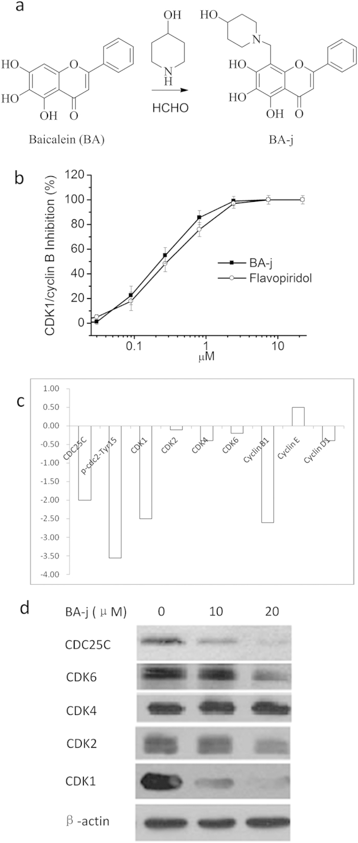
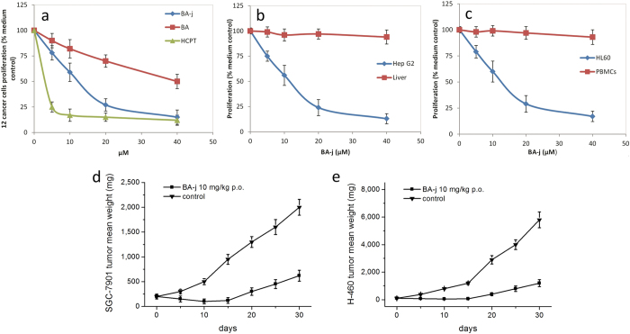
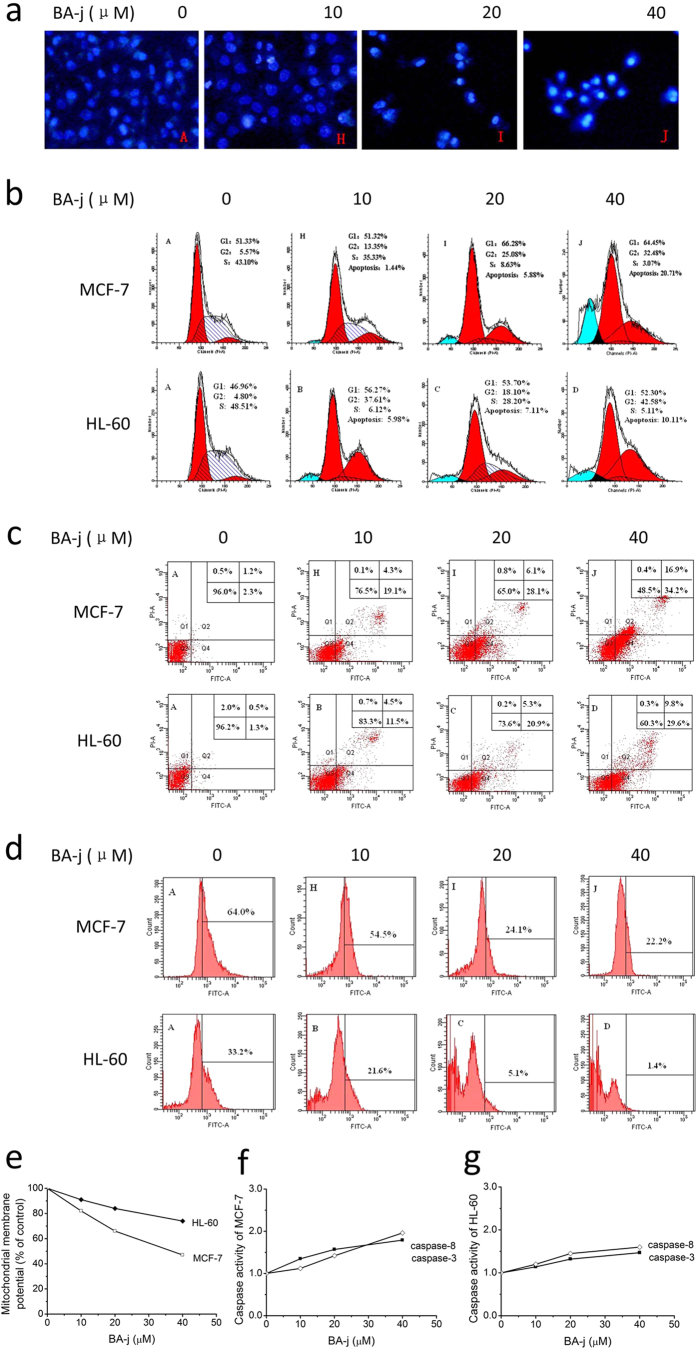
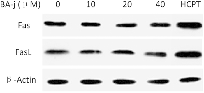
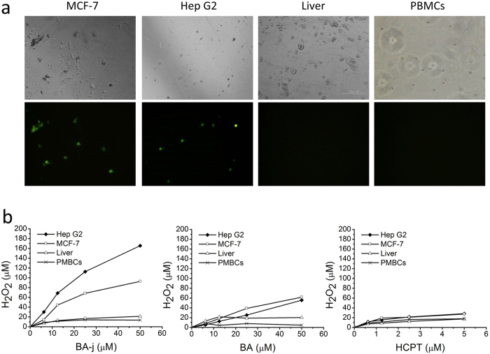
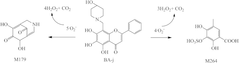
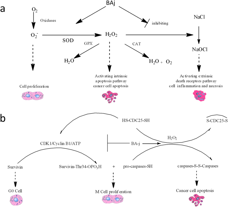
Cyclin-dependent kinase 1 (CDK1) is the only necessary CDK in cell proliferation and a novel target in the development of anticancer drugs. 8-Hydroxypiperidinemethyl-baicalein (BA-j) is a novel selective CDK1 inhibitor with broad spectrum anti-cancer activity (IC50 12.3 μM) and 2 tumor xenografts. Because of the differential mechanisms controlling redox-states in normal and cancer cells, BA-j can capture oxygen free radicals ((·)O2(-)) and selectively increase the level of H2O2 in cancer cells, thereby specifically oxidize and activate the intrinsic apoptosis pathway bypassing the extrinsic death receptor pathway, thus inducing apoptosis in cancer cells rather than in normal cells. BA-j is different from cytotoxic anticancer drugs which can activate both the intrinsic apoptosis pathway and the extrinsic death receptor pathway, and therefore harm normal cells while killing cancer cells. The molecular and biochemical mechanisms of reactive oxygen species (ROS) regulation suggest that BA-j may be developed into a novel anticancer agent.


Figures

Figure 1
Figure 1. Preparation and Targets screening of 8-Hydroxypiperidinemethyl-baicalein (BA-j).
(a) Preparation of BA-j. (b) Inhibitory activity against CDK1 of 8-Hydroxypiperidinemethyl-baicalein (BA-j). (c) Hep G2 cells were treated with 10 μM BA-j for 24 h, and the assay of protein level in HepG2 cell signaling pathways were determined by Western blot for conjugated secondary antibody (goat-antimouse IgG/FITC) using densitometric scanning and normalized using internal standards (β-actin). (d) Hep G2 cells were treated with 10 and 20 μM BA-j for 24 h, and the expressions of CDKs and CDC25C proteins in HepG2 cell signaling pathways were determined by Western blot for the appropriate secondary antibody (horseradish peroxidase-conjugated goat-antimouse IgG).
Figure 2
Figure 2. BA-j Anti-proliferation Activity in human cancer cells and 2 tumor xenografts.
Cells were cultured in the presence of BA-j for 48 h and an MTT assay was used to determine the anti-proliferation activity. (a) Anti-proliferation activity of BA-j in 12 human cancer cell lines compared with BA and HCPT treatment. (b) BA-j anti-proliferation activity comparison between Hep G2 cells and normal Liver cells. (c) BA-j anti-proliferation activity comparison between MCF-7 cells and normal PBMCs. (d,e) Tumor xenograft models of SGC-7901 and H-460 cells in male BALB/c nude mice treated with p.o. 10 mg/kg BA-j twice daily for 10 days.
Figure 3
Figure 3. Apoptosis Effects Assays of BA-j.
(a) Fluorescent images of Hoechst 33258 stained MCF-7 cells showing the induction of apoptosis following a 48 h BA-j treatment (200×). (b) Effects of BA-j treatment (48 h) on the cell cycle in MCF-7 and HL-60 cells were determined using PI staining. (c) Treating MCF-7 and HL-60 for 48 h with BA-j induces apoptosis. Cell apoptosis was demonstrated using the Annexin V-FITC and PI method. (d) Inhibition of Bcl-2 expression after a 48 h BA-j treatment in MCF-7 and HL-60 cells. (eg) The MMP is decreased and caspases are activated in MCF-7 and HL-60 cells treated with BA-j for 48 h. (e) BA-j treatment decreased the MMP in MCF-7 and HL-60 cells. (f) BA-j treatment activated caspase-8 and caspase-3 in MCF-7 cells. (g) BA-j treatment activated caspase-8 and caspase-3 in HL-60 cells.
Figure 4
Figure 4. Expression of Fas/FasL in Hep G2 cells treated with BA-j for 48 h compared with HCPT treatment.
2 μM HCPT was used.
Figure 5
Figure 5. Comparison of BA-j effects on cancer cells and normal cells.
(a) Fluorescent imaging of intracellular H2O2 (detected by PF1) in Hep G2 and MCF-7 cancer cells compared with normal Liver cells and PBMCs. All cells were treated with 12.5 μM BA-j for 4 h. (Top: bright field, Bottom: excitation wavelength 488 nm, emission wavelength 525 nm, 64×). (b) Assay of intracellular H2O2 following drug treatment in Hep G2 and MCF-7 cancer cells compared with normal Liver cells and PBMCs. The cells were cultured in the presence of drug for 4 h and the level of intracellular H2O2 was detected by PF1.
Figure 6
Figure 6. The major metabolic pathway of BA-j in macaques.
Figure 7
Figure 7. Mechanisms of ROS regulations.
(a) Molecular mechanisms of H2O2–regulated cell proliferation and apoptosis. (b) Mechanisms of ROS regulation by BA-j. BA-j can selectively induce apoptosis in cancer cells and protect normal cells from inflammation and necrosis.

Similar articles

Cited by

References

    1. Santamaria D. et al. Cdk1 is sufficient to drive the mammalian cell cycle. Nature 448, 811–815 (2007). - PubMed
    1. Malumbres M. & Barbacid M. Cell cycle, CDKs and cancer: a changing paradigm. Nat Rev Cancer 9, 153–166 (2009). - PubMed
    1. Diaz-Padilla I., Siu L. L. & Duran I. Cyclin-dependent kinase inhibitors as potential targeted anticancer agents. Invest New Drug 27, 586–594 (2009). - PubMed
    1. Rizzolio F., Tuccinardi T., Caligiuri I., Lucchetti C. & Giordano A. CDK inhibitors: from the bench to clinical trials. Curr Drug Targets 11, 279–290 (2010). - PubMed
    1. Wesierska-Gadek J., Maurer M., Zulehner N. & Komina O. Whether to target single or multiple CDKs for therapy? That is the question. J Cell Physiol 226, 341–349 (2011). - PubMed

Publication types

MeSH terms

LinkOut - more resources