Review
. 2015 Sep 1;7(9):a021683.
doi: 10.1101/cshperspect.a021683.

Motor Learning and the Cerebellum

Affiliations
Review

Motor Learning and the Cerebellum

Chris I De Zeeuw et al. Cold Spring Harb Perspect Biol. .

Abstract

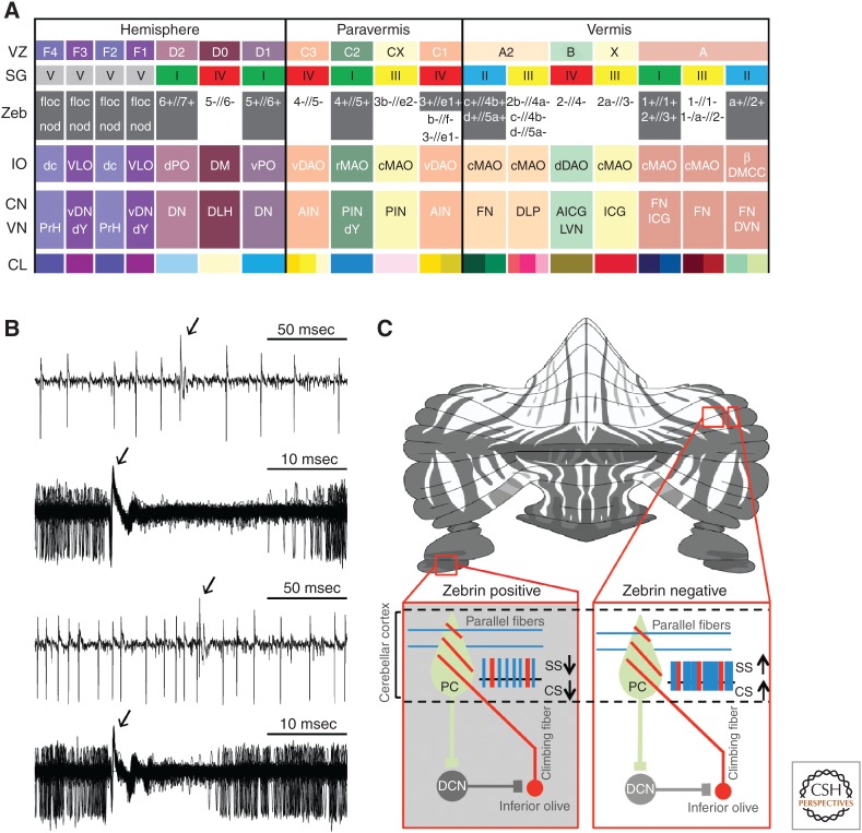
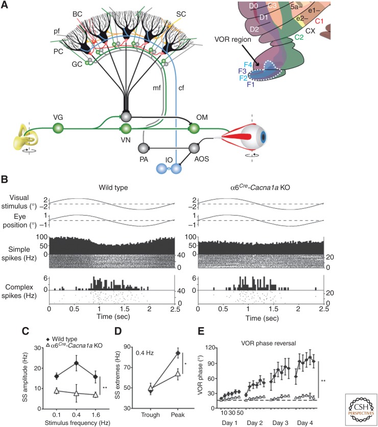
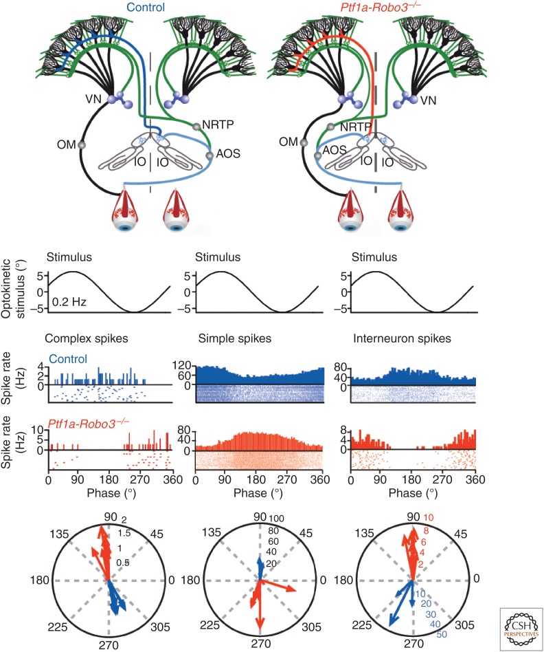
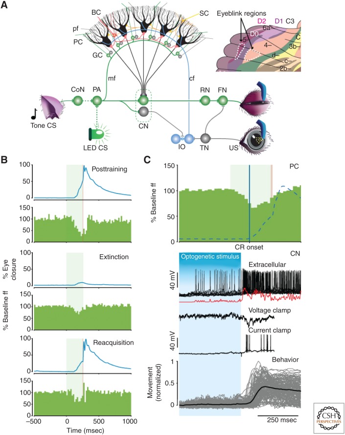
Although our ability to store semantic declarative information can nowadays be readily surpassed by that of simple personal computers, our ability to learn and express procedural memories still outperforms that of supercomputers controlling the most advanced robots. To a large extent, our procedural memories are formed in the cerebellum, which embodies more than two-thirds of all neurons in our brain. In this review, we will focus on the emerging view that different modules of the cerebellum use different encoding schemes to form and express their respective memories. More specifically, zebrin-positive zones in the cerebellum, such as those controlling adaptation of the vestibulo-ocular reflex, appear to predominantly form their memories by potentiation mechanisms and express their memories via rate coding, whereas zebrin-negative zones, such as those controlling eyeblink conditioning, appear to predominantly form their memories by suppression mechanisms and express their memories in part by temporal coding using rebound bursting. Together, the different types of modules offer a rich repertoire to acquire and control sensorimotor processes with specific challenges in the spatiotemporal domain.


Figures

Figure 1.
Figure 1.
Original identification of Purkinje cell zones in the cerebellar cortex of ferrets using Haggquist staining (based on data from Voogd 1964).
Figure 2.
Figure 2.
Olivocerebellar modules in mammals. The three-element modules of the olivocerebellar system are formed by a sagittal strip of Purkinje cells in the cerebellar cortex (A), which converge onto a particular set of cerebellar and/or vestibular nuclei (B–D), which, in turn, innervate the subnucleus in the inferior olive (E,F) that provides the climbing fibers to the corresponding strip of Purkinje cells, forming a closed triangular loop. (A) The left part of the cerebellar cortex indicates the original zones described by Voogd (1964), whereas the right part indicates the zebrin related groups described by Sugihara (2011). The color coding used in panels BF is the same as that used for describing Sugihara’s groups in A (i.e., right half), and together they reflect which parts within a particular olivocerebellar module are connected. For reference, we indicated the zebrin-positive strips with dark shading in Voogd’s zones on the left. (A) 1–6(a/b/–/+) (see Sugihara and Shinoda 2004, 2007); I–X, lobules I–X; CP, copula pyramidis; Cr I/II, crus I/II of ansiform lobule; FL, flocculus; Par, paramedian lobule; PFL, paraflocculus; Sim, lobulus simplex. (B,C) AICG, anterior interstitial cell group; AIN, anterior interposed nucleus; CP, copula pyramidis; DLH, dorsolateral hump; DLP, dorsolateral protuberance; DMC, dorsomedial crest; (v)DN, (ventral) dentate nucleus; FN, fastigial nucleus; ICG, interstitial cell group; PIN, posterior interposed nucleus. (D) DVN, descending vestibular nucleus; dY, dorsal group Y; MVN, medial vestibular nucleus; PrH, prepositus hypoglossal nucleus; SVN, superior vestibular nucleus. (E,F) β, subnucleus β; (c/v)DAO, (central/ventral) dorsal accessory olive; dc, dorsal cap; DM, dorsomedial group; DMCC, dorsomedial cell column; MAO, medial accessory olive; (d/v)PO, (dorsal/ventral) principal olive; VLO, ventrolateral outgrowth. Note that the X/CX-zones have only been found at the electrophysiological level (Ekerot and Larson 1982).
Figure 3.
Figure 3.
Olivocerebellar modules and Purkinje cell activity in relation to zebrin (II) distribution. (A) The VZ and SG rows refer to the zones and groups of Purkinje cells described by Voogd (1964; VZ) and Sugihara (2011; SG), respectively. The Zeb row indicates which zones and groups are zebrin positive (grey) and zebrin negative (white). The inferior olive (IO) row indicates which subnucleus of the IO is providing climbing fibers to a particular zone/group of Purkinje cells in the cerebellar cortex and collaterals to a particular part of the cerebellar nucleus, depicted in the same column. The CN and VN row indicates the parts of the cerebellar nuclei (CN) and vestibular nuclei (VN) that are innervated by the strip of Purkinje cells, depicted in the same column. It should be noted that only those vestibular nuclei are indicated that both receive a Purkinje cell input and provide a feedback projection to the inferior olive; because, for example, medial and superior vestibular nuclei do not project to the IO, they are not incorporated in this scheme. In addition, it should be noted that this overview is also incomplete in that some nuclei, such as the dorsomedial cell column (DMCC), may receive inhibitory feedback from multiple hindbrain regions. CL indicates the color legends used for Figure 2. AICG, anterior interstitial cell group; AIN, anterior interposed nucleus; β, subnucleus β; c/rMAO, caudal/rostral medial accessory olive; dc, dorsal cap; DLH, dorsolateral hump; DLP, dorsolateral protuberance; DM, dorsomedial group; DMC, dorsomedial crest; d/vDAO, dorsal/ventral dorsal accessory olive; DVN, descending vestibular nucleus; d/vPO, dorsal/ventral principal olive; dY, dorsal group Y; floc, flocculus; FN, fastigial nucleus; ICG, interstitial cell group; LVN, lateral vestibular nucleus; nod, nodulus; PIN, posterior interposed nucleus; PrH, prepositus hypoglossal nucleus; vDN, ventral dentate nucleus; and VLO, ventrolateral outgrowth. (B) Examples of raw traces of Purkinje cell activity from zebrin-positive (top panels) and zebrin-negative (bottom panels) zones. Arrows indicate complex spike. (From Zhou et al. 2014; reprinted, with permission, from the authors.) (C) Intramodular connections via deep cerebellar nuclei (DCN) explaining why the complex spike (CS) activity within a module follows the intrinsic differences in simple spike (SS) activity of Purkinje cells (PC). PC and DCN are inhibitory, whereas climbing fibers are excitatory. (From Albergaria and Carey 2014; reprinted under the terms of the Creative Commons Attribution License, which permits unrestricted use and redistribution provided that the original author and source are credited.)
Figure 4.
Figure 4.
Circuit of the vestibulo-ocular reflex and prominent role of modulation amplitude. (A) The vestibulo-ocular reflex (VOR) is mediated by the three-neuron arc of Lorente de No in the brainstem. When the head rotates, the vestibular signals from the semicircular canals are transferred by the vestibular ganglion cells (VG) to the second-order vestibular neurons in the vestibular nuclei (VN), which in turn innervate the oculomotor neurons (OMs) driving the eyes to the opposite side. The vestibulocerebellum, which is superimposed on this three-neuron arc, is required to compensate for the delays introduced during input–output processing. To minimize retinal slip during head movements, the accessory optic system (AOS) relays slip signals through the climbing fiber (cf) system to the zebrin-positive Purkinje cells (PC) in floccular zones F1–F4 in the vestibulocerebellar cortex (see inset), where the presence and absence of the climbing fiber activity is integrated with vestibular, optokinetic, and eye movement signals mediated by the mossy fiber (mf)–granule cell (GC)–parallel fiber (pf) pathway. The Purkinje cells in turn can inject well-calibrated, accelerating signals into the vestibular brainstem so as to precisely compensate for the delays. (B) α6Cre-Cacna1a knockout (KO) mice, in which transmission in the vast majority, but not all, of parallel fibers is blocked, show a normal amplitude of their optokinetic reflex when a visual stimulus is given, despite a reduced modulation of their simple spike activity. (C,D) The modulation of the simple spike, but not the complex spike, activity in α6Cre-Cacna1a KO is reduced over a wide range of frequencies, and these deficiencies are caused by a reduction in the peak of the modulation. (E) VOR phase reversal is a form of VOR adaptation, during which the phase of the VOR is reversed by providing an in-phase optokinetic stimulus that is greater in amplitude than the vestibular stimulus; α6Cre-Cacna1a KO mice have severe problems reversing the phase of their eye movements indicating that mammals have their abundance of GCs and pfs to control motor learning rather than basic motor performance. PA, pontine area; BC, basket cell; SC, stellate cell. (From Galliano et al. 2013a; modified, with permission, from the authors.)
Figure 5.
Figure 5.
Climbing fibers dominate timing of simple spike firing. Although the complex spikes of Purkinje cells are modulated by activity in the climbing fiber system, the simple spikes are supposed to be largely driven by the mossy fiber system. The frequencies of these two types of spikes are often modulated reciprocally. An increase in complex spikes is associated with a decrease in simple spikes, and vice versa. This reciprocal firing is thought to be essential for motor behavior. Rerouting the climbing fiber system in Ptf1a-Robo3 mice from a contralateral (dark blue line in top panel) to a predominantly ipsilateral projection (red line in top panel) does not only reverse the modulation of complex spike activity during natural optokinetic stimulation (see peristimulus–time histograms (PSTHs), raster, and polar plots in left panel), but also that of the simple spike activity (middle panel). Because the laterality of the mossy fiber projection is unaffected (green lines in top panels), these data show that the proper timing of the climbing fiber input is essential for well-coordinated motor performance by controlling the timing of simple spike firing. The phase of molecular layer interneurons is also reversed in the mutants (right panel), which suggests that climbing fibers evoke their effects on simple spike activity via molecular layer interneurons. VN, Vestibular nuclei; NRTP, nucleus reticularis tegmenti pontis; OM, oculomotor neuron; IO, inferior olive; AOS, accessory optic system. (From Badura et al. 2013; modified, with permission, from the authors.)
Figure 6.
Figure 6.
Eyeblink circuit and role of simple spike suppression. (A) During eyeblink conditioning, a conditioned stimulus (CS), such as a tone or LED light, is repetitively paired with an unconditioned stimuli (US), such as an airpuff, to learn a well-timed conditioned response (CR). CS and US sensory information converges at zebrin-negative Purkinje cells (PCs) in D0 and C3 (see inset) through the mossy fiber (mf)–granule cell (GC)–parallel fiber (pf) pathway and the climbing fibers (cfs) derived from the inferior olive (IO), respectively. Although the mossy fibers relay information on the CS from the pontine area (PA), the climbing fibers mediate efferent copies of signals evoked in the direct eyeblink reflex loop, which is formed by the orbital branch of the trigeminal nerve, trigeminal nucleus (TN), facial nucleus (FN), and eyelid muscle. When a fixed temporal relationship between parallel fiber and climbing fiber activation emerges, the same parallel fiber input starts to evoke a simple spike suppression that disinhibits the cerebellar nuclear (CN) cells, and consequently causes the eyelid to close before the US is about to occur. (B) Example of mean eyelid behavioral traces and simple spike frequency histograms of a mouse Purkinje cell from lobule HVI (zebrin-negative D0 zone) after training, extinction, and reacquisition (top to bottom). Note the concomitant changes in simple spike suppression and amplitude of the CRs. The green and red bands in the background depict CS and US duration, respectively. (C) (Top) Simple spike suppression precedes CR onset (blue line) and covaries with its course (dashed line). (Bottom) A rapid drop in Purkinje cell activity after stopping optogenetic stimulation elicits rebound burst activity in CN neurons (extracellular recording in vivo). The excitatory events in voltage and current clamp recordings of the same cell in vivo following optogenetic Purkinje cell stimulation show that this bursting may be facilitated by climbing and/or mossy fiber collaterals. Ultimately, CN rebound activity can reliably evoke a behavioral response. The data described under C are obtained from different experiments, but aligned at the same time scale to facilitate understanding of the course of events with respect to each other. CoN, cochlear nucleus; RN, red nucleus. (From Witter et al. 2013; modified, with permission, from the authors.)

References

    1. Albergaria C, Carey MR. 2014. All Purkinje cells are not created equal. eLife 3: e03285. - PMC - PubMed
    1. Albus JS. 1971. A theory of cerebellar function. Math Biosci 10: 25–61.
    1. Andreescu CE, Milojkovic BA, Haasdijk ED, Kramer P, De Jong FH, Krust A, De Zeeuw CI, De Jeu MT. 2007. Estradiol improves cerebellar memory formation by activating estrogen receptor β. J Neurosci 27: 10832–10839. - PMC - PubMed
    1. Angaut P, Compoint C, Buisseret-Delmas C, Batini C. 1996. Synaptic connections of Purkinje cell axons with nucleocortical neurones in the cerebellar medial nucleus of the rat. Neurosci Res 26: 345–348. - PubMed
    1. Apps R, Hawkes R. 2009. Cerebellar cortical organization: A one-map hypothesis. Nat Rev Neurosci 10: 670–681. - PubMed

Publication types

LinkOut - more resources