. 2015 Oct 15;128(20):3744-56.
doi: 10.1242/jcs.172536. Epub 2015 Sep 1.

Failure of lysosome clustering and positioning in the juxtanuclear region in cells deficient in rapsyn

Affiliations

Failure of lysosome clustering and positioning in the juxtanuclear region in cells deficient in rapsyn

Mohamed Aittaleb et al. J Cell Sci. .

Abstract

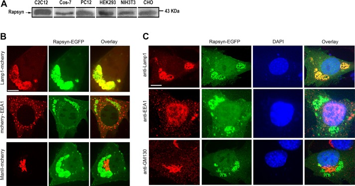
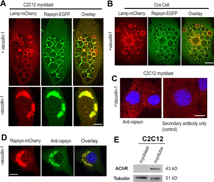
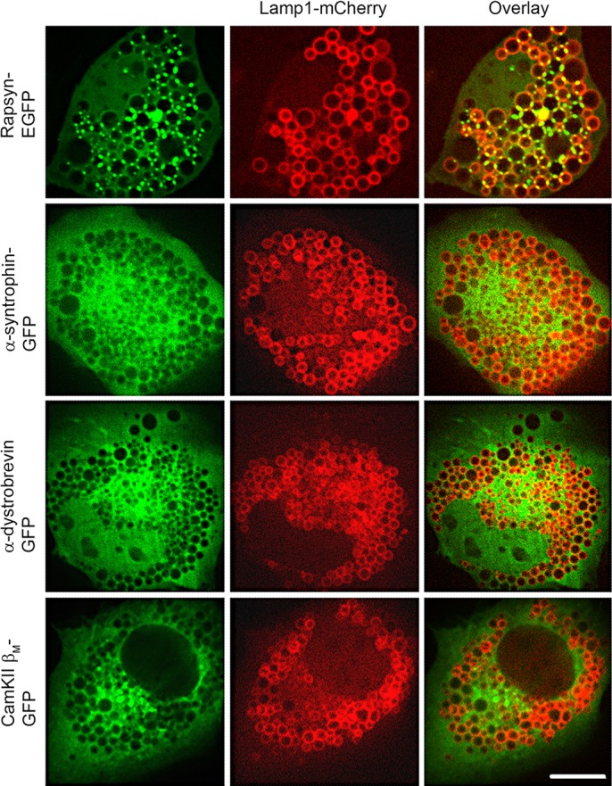
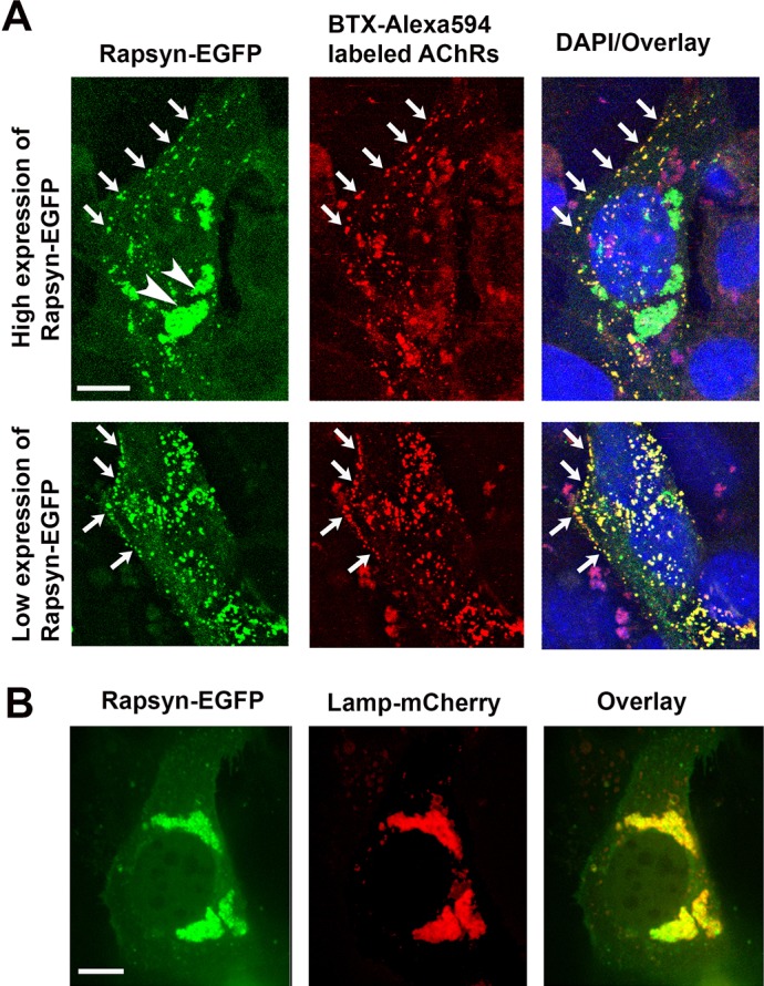
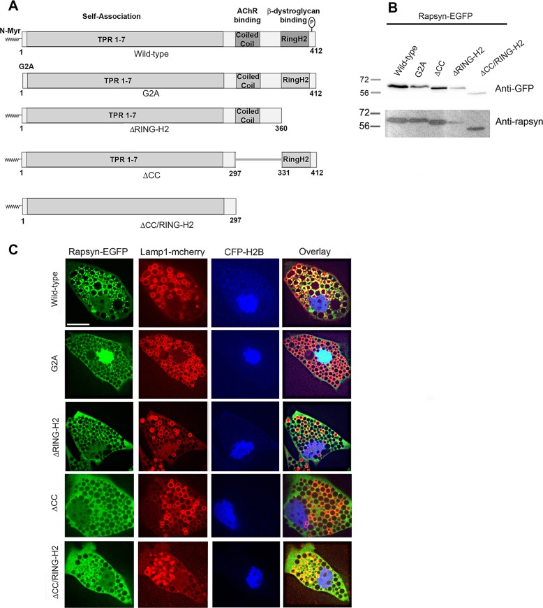
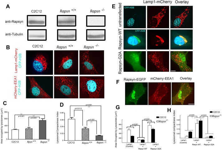
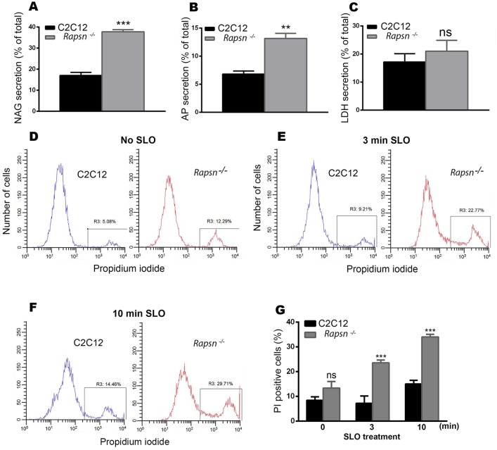
Rapsyn, a scaffold protein, is required for the clustering of acetylcholine receptors (AChRs) at contacts between motor neurons and differentiating muscle cells. Rapsyn is also expressed in cells that do not express AChRs. However, its function in these cells remains unknown. Here, we show that rapsyn plays an AChR-independent role in organizing the distribution and mobility of lysosomes. In cells devoid of AChRs, rapsyn selectively induces the clustering of lysosomes at high density in the juxtanuclear region without affecting the distribution of other intracellular organelles. However, when the same cells overexpress AChRs, rapsyn is recruited away from lysosomes to colocalize with AChR clusters on the cell surface. In rapsyn-deficient (Rapsn(-/-)) myoblasts or cells overexpressing rapsyn mutants, lysosomes are scattered within the cell and highly dynamic. The increased mobility of lysosomes in Rapsn(-/-) cells is associated with a significant increase in lysosomal exocytosis, as evidenced by increased release of lysosomal enzymes and plasma membrane damage when cells were challenged with the bacterial pore-forming toxin streptolysin-O. These findings uncover a new link between rapsyn, lysosome positioning, exocytosis and plasma membrane integrity.

Keywords: Clustering; Exocytosis; Lysosomes; Mobility; Rapsyn.


Conflict of interest statement

Competing interests

The authors declare no competing or financial interests.

Figures

Fig. 1.
Fig. 1.
Rapsyn expression and localization in non-muscle and undifferentiated myoblast cells. (A) Immunoblots of lysates from undifferentiated C2C12 myoblasts and non-muscle cell types (COS-7, PC12, HEK293, NIH3T3 and CHO) probed with rabbit monoclonal anti-rapsyn antibody. Note that rapsyn is present in extracts of all examined cells. (B) Undifferentiated C2C12 myoblasts were co-transfected with rapsyn–EGFP and either the lysosomal marker Lamp1–mCherry, early endosome marker mCherry–EEA1 or Golgi marker ManII–mCherry and then cells expressing fluorescent fusion proteins were live imaged using the confocal spinning disk microscope. In the top left panel representative images of live C2C12 myoblasts co-transfected with rapsyn–EGFP and lysosomal marker Lamp1–mCherry are shown; in the middle left panel rapsyn–EGFP and early endosome marker mCherry–EEA1 are shown; in the bottom left panel rapsyn–EGFP and Golgi marker ManII–mCherry are shown. Note that rapsyn colocalizes only with lysosomes. (C) Immunostaining of C2C12 myoblasts expressing rapsyn–EGFP with rat anti-Lamp1, rabbit anti-EEA1 or mouse anti-GM130 antibodies. Note that rapsyn localizes to lysosomes but not to endosomes or Golgi. Scale bars: 10 µm.
Fig. 2.
Fig. 2.
Rapsyn concentrates at junctional sites between vacuolin-1 enlarged lysosomal vacuoles. (A) Undifferentiated C2C12 myoblasts or (B) non-muscle COS-7 cells were transfected with rapsyn–EGFP and Lamp1–mCherry and either left untreated (lower panels) or treated with vacuolin-1 (upper panels). Rapsyn–EGFP colocalizes perfectly with Lamp1–mCherry in the juxtanuclear region of non-treated C2C12 myoblasts (A, lower panels) and is concentrated at the junctional sites between enlarged lysosomal vacuoles visualized by Lamp1–mCherry (A, upper panels) and in COS cells (B). (C) C2C12 myoblasts were treated with vacuolin-1, fixed and labeled with mouse monoclonal anti-rapsyn antibody followed by a secondary fluorescent antibody (left panel) or with a fluorescent secondary antibody only (right panel). (D) C2C12 myoblast transfected with rapsyn–mCherry, fixed and labeled with antibody against rapsyn. Scale bars: 10 µm. (E) Blot showing that AChRs are undetectable in undifferentiated myoblasts.
Fig. 3.
Fig. 3.
Rapsyn, but not other proteins of the postsynaptic complex, is selectively targeted to lysosomes. C2C12 myoblasts were co-transfected with Lamp1–mCherry and either rapsyn–EGFP, α-syntrophin–GFP, α-dystrobrevin–GFP or CamKIIβM–GFP. Cells were then treated with vacuolin-1 prior to live imaging by confocal spinning disk microscope. Representative images showing that rapsyn–EGFP accumulates selectively at the junctional sites between enlarged lysosomal vacuoles whereas α-syntrophin–GFP, α-dystrobrevin–GFP or CamKIIβM–GFP showed a diffuse distribution throughout the cytoplasm, with no localization to the vacuoles. Scale bars: 10 µm.
Fig. 4.
Fig. 4.
Rapsyn is recruited to the cell surface in the presence of AChRs. C2C12 myoblasts were co-transfected with rapsyn–EGFP and either (A) AChRα, AChRβ, AChRδ and AChRε subunits or (B) Lamp1–mCherry. (A) In C2C12 myoblasts expressing AChRs and high levels of rapsyn–EGFP, rapsyn is concentrated in the juxtanuclear region (arrowheads) and colocalizes with AChR clusters at the cell surface (upper panel). However, in myoblast expressing AChRs and low level of rapsyn, most rapsyn molecules are recruited to AChR clusters at the cell surface (lower panel). Arrows indicate co-clusters of AChRs and rapsyn at the cell surface. (B) When myoblasts were transfected with rapsyn–EGFP and Lamp1–mCherry, rapsyn remains largely concentrated with Lamp1–mCherry in the juxtanuclear region (no visible accumulation at the cell surface). Scale bars: 10 µm.
Fig. 5.
Fig. 5.
The targeting of rapsyn to the junctional sites between lysosomal vacuoles requires the myristoylation, coiled-coil and RING-H2 domains. (A) Schematic representation of rapsyn constructs used as EGFP fusions in this study. (B) Lysates from COS cells expressing the indicated rapsyn–EGFP construct were blotted with anti-GFP or anti-rapsyn antibody and showed the corresponding proteins at expected sizes confirming the intact protein fusions. (C) C2C12 myoblasts were co-transfected with Lamp1–mCherry (a lysosomal marker), CFP-H2B (a nuclear marker) and either wild-type or the indicated rapsyn–EGFP mutant and then treated with vacuolin-1. Shown are representative images of cells transfected with Lamp1–mCherry and the indicated rapsyn–EGFP construct. Note that non-myristoylated rapsyn G2A–EGFP is concentrated in the nucleus. Rapsyn ΔCC–EGFP, rapsyn ΔRING-H2–EGFP and rapsyn ΔCC/RING-H2–EGFP are more diffuse in the cytoplasm whereas wild-type rapsyn–EGFP is concentrated at junctional sites between enlarged lysosomes. Scale bar: 10 µm.
Fig. 6.
Fig. 6.
Overexpression of rapsyn mutants induces the declustering of lysosomes and disruption of microtubules had no effect on rapsyn and lysosomes clusters in the juxtanuclear region. C2C12 myoblasts were transfected with Lamp1–mCherry (a lysosomal marker) and CFP–H2B to label the nuclei (control) or co-transfected with Lamp1–mCherry and either wild-type, G2A, ΔCC, ΔRING-H2 or ΔCC/RING-H2 rapsyn–EGFP constructs. CFP–H2B was only used when myoblasts were transfected with Lamp1–mCherry alone to better visualize the nucleus. Transfected myoblasts were then imaged live using confocal spinning disk microscopy. (A) Representative images of cells expressing the lysosomal marker Lamp1–mCherry and CFP–H2B (control) or Lamp1–mCherry and the indicated form of rapsyn–EGFP. Note that the clustering of lysosomes in the juxtanuclear region was significantly increased in presence of wild-type rapsyn–EGFP, whereas rapsyn mutant constructs induced the declustering of lysosomes and their scattering throughout the cytoplasm. (B) Graph showing the area occupied by lysosomes in µm2. Note that in the presence of wild-type rapsyn, the area occupied by lysosomes is decreased compared to control cells, whereas this area is significantly increased in the presence of each rapsyn mutant. (C) Graph showing quantification of the lysosomal compactness index. Note that compactness index significantly increased in the presence of wild-type rapsyn, but dramatically decreased in the presence of rapsyn mutants compared to untransfected cells. The compactness index was determined as described in the Materials and Methods and as reported previously (Bard et al., 2003; Zilberman et al., 2011). The P-values were calculated using a Bonferroni test. Results are mean±s.e.m. (D) Myoblasts were transfected with rapsyn–mCherry and EMTB–3×EGFP or rapsyn–EGFP and Lamp1–mCherry and then treated with nocodazole, a drug that disrupts microtubules. Upper panels show examples of myoblast cells untreated or treated with nocodazole that were live imaged using a confocal spinning disk. The lower panel shows an example of a myoblast that was treated with nocodazole, fixed and imaged. Note that in the presence of nocodazole, rapsyn and lysosomes clusters remained intact in the juxtanuclear region whereas the microtubule-binding domain of ensconsin–EGFP was completely disrupted. Scale bars: 10 µm.
Fig. 7.
Fig. 7.
The clustering of lysosomes in the juxtanuclear region is impaired in rapsyn-deficient myoblasts, whereas exogenous wild-type rapsyn, but not its non-myristoylated form, rescues the lysosome clustering phenotype. (A) Lysates from C2C12, Rapsn+/+ or Rapsn−/− myoblasts were blotted for endogenous rapsyn using a rabbit monoclonal anti-rapsyn antibody or mouse anti-tubulin for loading control. Endogenous rapsyn was detected in C2C12 myoblasts and Rapsn+/+ myoblasts but not in Rapsn−/− myoblasts. (B) Confocal images of live myoblasts expressing CFP–H2B (to label nuclei) and either Lamp1–mCherry or mCherry–EEA1. The top panel shows representative images of C2C12, Rapsn+/+ or Rapsn−/− myoblasts transfected with lysosomal marker Lamp1–mCherry and CFP–H2B. Although lysosomes are distributed in the juxtanuclear region of C2C12 and Rapsn+/+ myoblasts, they are instead scattered through the entire cytosol in Rapsn−/− myoblasts. The bottom panel shows cells expressing EEA1–mCherry and CFP–H2B. Note that the localization of endosomes is not affected by the loss of endogenous rapsyn as a similar distribution of EEA1 was observed in C2C12, Rapsn+/+ and Rapsn−/− myoblasts, indicating that rapsyn selectively affects the lysosomes clustering. (C) Quantification of the area occupied by lysosomes showing a 2-fold increase in the mean area in Rapsn−/− compared to either Rapsn+/+ or C2C12 myoblasts. P<0.001. Results are mean±s.e.m. (D) Graph showing the quantification of lysosomal compactness index. Note that the mean compactness index in Rapsn−/− is reduced 2.6 times compared to Rapsn+/+ and 4.6 times compared to C2C12 myoblasts. The P values were calculated using Bonferroni test. Results are mean±s.e.m. (E) Representative images of Rapsn−/− myoblasts expressing CFP–H2B (to label nuclei) and Lamp1–mCherry without (top panel), or with wild-type rapsyn–EGFP (middle panel), or Lamp1–mCherry and rapsynG2A–EGFP (lower panel). Note that the scattering of the lysosomes in the absence of rapsyn (top panel) was completely rescued in the presence of exogenous wild-type rapsyn–EGFP as the lysosomes clustered back into the juxtanuclear region. In contrast, expression of rapsynG2A–EGFP was unable to reverse the lysosome scattering defect of Rapsn−/− myoblasts and the lysosomes remained scattered throughout the cytoplasm (lower panel). (F) Representative images of cells showing that the scattered distribution of early endosomes was not affected by overexpression of wild-type rapsyn–EGFP. Scale bars: 10 µm. (G) Quantification of the area occupied by lysosomes in Rapsn−/− myoblasts transfected with Lamp1–mCherry only (untransfected control) or with wild-type rapsyn–EGFP or rapsynG2A–EGFP. Note that in the presence of wild-type rapsyn–EGFP, the area occupied by lysosomes is 5-fold lower than in the absence of rapsyn–EGFP and 6.7-fold lower than in the presence of rapsynG2A–EGFP. For comparison, corresponding data for C2C12 myoblasts are shown side by side. Note that wild-type rapsyn–EGFP rescues the lysosome clustering to the level observed in C2C12 myoblasts. Results are mean±s.e.m. (H) Graph summarizing the quantification of the lysosome compactness index. Note that the index of lysosomal compactness is significantly increased in Rapsn−/− myoblasts transfected with exogenous wild-type rapsyn–EGFP (0.16±0.02, mean±s.e.m., n=10) compared to control Rapsn−/− myoblasts (0.014±0.001, mean±s.e.m., n=29) or Rapsn−/− myoblasts transfected with rapsynG2A–EGFP (0.008±0.001, mean±s.e.m., n=13).
Fig. 8.
Fig. 8.
Lysosomal exocytosis was significantly increased in Rapsn−/− myoblasts. Graphs showing lysosomal enzyme activities. (A) β-N-acetylglucoseaminidase (NAG), (B) acid phosphatase (AP) and (C) Lactate dehydrogenase (LDH). Note that both lysosomal enzymes were significantly increased in rapsyn-deficient myoblasts compared to C2C12 myoblasts. Results are mean±s.e.m. (D–F) Representative histograms obtained by FACS analysis of propidium iodide staining of live cells: (D) non-treated C2C12 myoblasts (left) and Rapsn−/− (right) myoblasts; (E) treated (permeabilized) myoblasts exposed to 500 µg/ml of bacterial toxin streptolysin-O (SLO) for 3 min or (F) 10 min. In each histogram, the left peak corresponds to intact (propidium iodide negative) cells and the right peak corresponds to propidium-iodide-positive cells. Note that untreated Rapsn−/− myoblasts show a higher basal uptake of propidium iodide than C2C12 cells, indicative of plasma membrane damage. This difference in propidium iodide uptake becomes larger as cells are challenged with SLO for 3 min (E) or 10 min (F), indicating that in the absence of rapsyn, plasma membrane is more vulnerable to damage. (G) Quantification of FACS data (as shown in D,E, and F), which is reported as percentage of propidium-iodide-positive Rapsn−/− and C2C12 myoblasts that were either untreated (0 min) or treated with SLO for the indicated periods of time. The data represent mean±s.e.m. from three independent experiments.

References

    1. Abenza J. F., Galindo A., Pantazopoulou A., Gil C., de los Rios V. and Penalva M. A. (2010). Aspergillus RabBRab5 integrates acquisition of degradative identity with the long distance movement of early endosomes. Mol. Biol. Cell 21, 2756-2769. 10.1091/mbc.E10-02-0119 - DOI - PMC - PubMed
    1. Abenza J. F., Galindo A., Pinar M., Pantazopoulou A., de los Rios V. and Penalva M. A. (2012). Endosomal maturation by Rab conversion in Aspergillus nidulans is coupled to dynein-mediated basipetal movement. Mol. Biol. Cell 23, 1889-1901. 10.1091/mbc.E11-11-0925 - DOI - PMC - PubMed
    1. Andrews N. W., Almeida P. E. and Corrotte M. (2014). Damage control: cellular mechanisms of plasma membrane repair. Trends Cell Biol. 24, 734-742. 10.1016/j.tcb.2014.07.008 - DOI - PMC - PubMed
    1. Antolik C., Catino D. H., O'Neill A. M., Resneck W. G., Ursitti J. A. and Bloch R. J. (2007). The actin binding domain of ACF7 binds directly to the tetratricopeptide repeat domains of rapsyn. Neuroscience 145, 56-65. 10.1016/j.neuroscience.2006.11.047 - DOI - PMC - PubMed
    1. Balderhaar H. J. k. and Ungermann C. (2013). CORVET and HOPS tethering complexes - coordinators of endosome and lysosome fusion. J. Cell Sci. 126, 1307-1316. 10.1242/jcs.107805 - DOI - PubMed

Publication types