. 2015 Nov;355(2):329-40.
doi: 10.1124/jpet.115.226969. Epub 2015 Sep 1.

Identification and characterization of modified antisense oligonucleotides targeting DMPK in mice and nonhuman primates for the treatment of myotonic dystrophy type 1

Affiliations

Identification and characterization of modified antisense oligonucleotides targeting DMPK in mice and nonhuman primates for the treatment of myotonic dystrophy type 1

Sanjay K Pandey et al. J Pharmacol Exp Ther. 2015 Nov.

Abstract

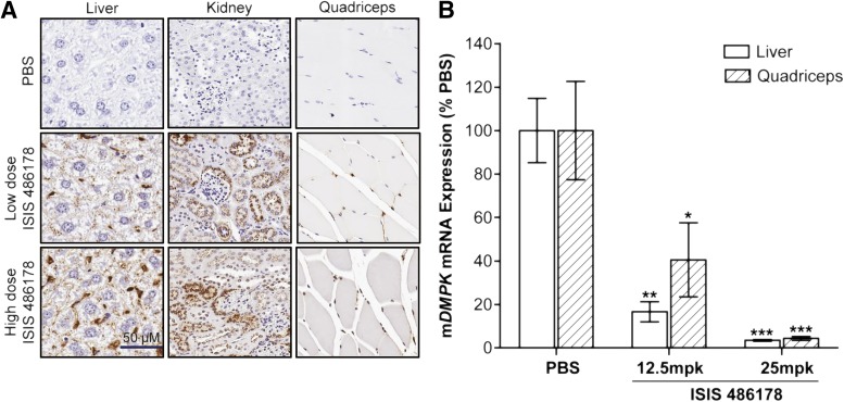
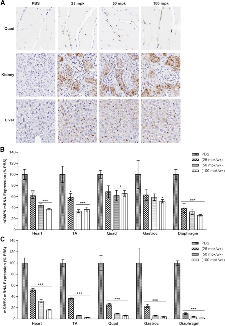
Myotonic dystrophy type 1 (DM1) is the most common form of muscular dystrophy in adults. DM1 is caused by an expanded CTG repeat in the 3'-untranslated region of DMPK, the gene encoding dystrophia myotonica protein kinase (DMPK). Antisense oligonucleotides (ASOs) containing 2',4'-constrained ethyl-modified (cEt) residues exhibit a significantly increased RNA binding affinity and in vivo potency relative to those modified with other 2'-chemistries, which we speculated could translate to enhanced activity in extrahepatic tissues, such as muscle. Here, we describe the design and characterization of a cEt gapmer DMPK ASO (ISIS 486178), with potent activity in vitro and in vivo against mouse, monkey, and human DMPK. Systemic delivery of unformulated ISIS 486718 to wild-type mice decreased DMPK mRNA levels by up to 90% in liver and skeletal muscle. Similarly, treatment of either human DMPK transgenic mice or cynomolgus monkeys with ISIS 486178 led to up to 70% inhibition of DMPK in multiple skeletal muscles and ∼50% in cardiac muscle in both species. Importantly, inhibition of DMPK was well tolerated and was not associated with any skeletal muscle or cardiac toxicity. Also interesting was the demonstration that the inhibition of DMPK mRNA levels in muscle was maintained for up to 16 and 13 weeks post-treatment in mice and monkeys, respectively. These results demonstrate that cEt-modified ASOs show potent activity in skeletal muscle, and that this attractive therapeutic approach warrants further clinical investigation to inhibit the gain-of-function toxic RNA underlying the pathogenesis of DM1.


Figures

Fig. 1.
Fig. 1.
Treatment with DMPK-targeting ISIS 486178 reduces DMPK mRNA in multiple human cell types and cynomolgus monkey hepatocytes. (A) Region of hDMPK targeted by ISIS 486178 relative to expanded CUG repeat in 3′-UTR. ISIS 486178 was electroporated into (B) HepG2 cells, (C) DM1/Steinert myoblasts (>1000 CTG repeats), and (D) cynomolgus monkey primary hepatocytes at the indicated concentrations. After 24 hours, cells were lysed and total RNA was isolated and human DMPK mRNA levels were determined by qPCR and normalized to total RNA. A control ASO was examined. Error bars represent standard errors of means (n = 2 to 3). ***P < 0.001 versus untreated control (UTC) using two-way analysis of variance. (E) Reduction of RNA foci in DM1 patient myoblasts upon treatment with DMPK ASO. DM1 myoblast cells were treated with ISIS 486178 at 20 nM concentration for 24–48 hours (bottom panels). Control treatment groups are shown in top panels. After the treatment, fluorescent in situ hybridization was performed on these cells. Nuclei were stained with DAPI (blue) and CUGexp RNA foci (red).
Fig. 2.
Fig. 2.
Localization and DMPK mRNA expression in tissues of wild-type mice after systemic administration of ISIS 486178. (A) ISIS 486178 was administered s.c. to normal C57Bl/6 mice (n = 4 per dose group) at 12.5 or 25 mg/kg body weight twice a week for 6 weeks. ASO levels in the liver, kidney, and quadriceps muscle were determined by immunostaining with anti-ASO antibody, followed by counterstaining with hematoxylin. (B) C57Bl/6 mice (n = 4 per dose group) were treated s.c. with PBS or ISIS 486178 twice weekly for 6 weeks at doses of 12.5 or 25 mg/kg body weight. Animals were sacrificed 48 hours after the final dose, and the liver and quadriceps muscle were collected. DMPK mRNA levels were measured by quantitative real-time PCR and normalized to total RNA. Error bars represent standard errors of means (n = 3 to 4). *P < 0.05, **P < 0.01, and ***P < 0.001 versus PBS-treated group using two-way analysis of variance, followed by Bonferonni multiple comparison tests.
Fig. 3.
Fig. 3.
ISIS 486178 treatment reduced both human and endogenous mouse DMPK mRNA expression in DMSXL mice expressing the human DMPK gene with >1000 CTG repeats. DMSXL mice (n = 5 per group) were injected s.c. with PBS or ISIS 486178 at doses of 12.5, 25, and 50 mg/kg body weight twice a week for 6 weeks. Mice were sacrificed 2 days after the last dose. Organs and blood samples were collected postsacrifice. (A) ISIS 486178 was visualized in the liver, kidney, and quadriceps muscle by immunostaining with anti-ASO antibody, followed by counterstaining with hematoxylin. (B) Human and (C) mouse DMPK mRNA levels were quantified by quantitative real-time PCR and normalized to total RNA input. Error bars represent standard errors of means. *P < 0.05, **P < 0.01, and ***P < 0.001 versus PBS-treated group using one-way analysis of variance, followed by Bonferonni post-tests.
Fig. 4.
Fig. 4.
Duration of action of ISIS 486178 in DMSXL mice. Four cohorts of 10- to 12-week-old DMSXL mice were injected subcutaneously with PBS or ISIS 486178 at a dose of 25 mg/kg of body weight twice weekly for 6 weeks (n = 5). Groups were sacrificed 2 days and 6, 15, and 31 weeks after the last dose. Heart, TA, quadriceps (Quad), gastrocnemius (Gastroc), and diaphragm tissues were harvested. RNA was isolated, and quantitative real-time PCR was performed to determine levels of (A) hDMPK and (B) mDMPK mRNAs. Error bars represent standard errors of means. *P < 0.05, **P < 0.01, and ***P < 0.001 versus PBS-treated groups using two-way analysis of variance, followed by Bonferonni multiple comparison test.
Fig. 5.
Fig. 5.
ISIS 486178 reduces DMPK mRNA expression in normal cynomolgus monkey tissues, with a prolonged duration of action in TA muscle. (A) Male monkeys (n = 4 per group) received s.c. injections of PBS or 40 mg/kg ISIS 486178 on days 1, 3, 5, and 7 and once weekly for another 12 weeks (13 weeks total). Several tissues were isolated 2 days after the last dose. Another group of monkeys dosed similarly was followed for 26 weeks. DMPK mRNA was measured by quantitative real-time PCR and normalized to total RNA levels. Quad, quadriceps; Gastroc, gastrocnemius. (B) Male monkeys (n = 4 per group) received s.c. injections of PBS or ISIS 486178 at days 1, 3, 5, and 7 and once weekly for another 12 weeks. TA muscle was biopsied at week 7 (during treatment) and week 19 (6 weeks post-treatment) as well as harvested at necropsy at week 13 (completion of the treatment) and 26 (13 weeks post-treatment). DMPK mRNA was measured by quantitative real-time PCR and normalized to total RNA levels. Error bars represent standard errors of means. *P < 0.05, **P < 0.01, and ***P < 0.001 versus PBS-treated groups using one-way analysis of variance, followed by Dunnett’s multiple comparison test.

Similar articles

Cited by

References

    1. Agrawal S, Zhao Q, Jiang Z, Oliver C, Giles H, Heath J, Serota D. (1997) Toxicologic effects of an oligodeoxynucleotide phosphorothioate and its analogs following intravenous administration in rats. Antisense Nucleic Acid Drug Dev 7:575–584. - PubMed
    1. Bennett CF, Swayze EE. (2010) RNA targeting therapeutics: molecular mechanisms of antisense oligonucleotides as a therapeutic platform. Annu Rev Pharmacol Toxicol 50:259–293. - PubMed
    1. Berul CI, Maguire CT, Aronovitz MJ, Greenwood J, Miller C, Gehrmann J, Housman D, Mendelsohn ME, Reddy S. (1999) DMPK dosage alterations result in atrioventricular conduction abnormalities in a mouse myotonic dystrophy model. J Clin Invest 103:R1–R7. - PMC - PubMed
    1. Berul CI, Maguire CT, Gehrmann J, Reddy S. (2000) Progressive atrioventricular conduction block in a mouse myotonic dystrophy model. J Interv Card Electrophysiol 4:351–358. - PubMed
    1. Blake DJ, Weir A, Newey SE, Davies KE. (2002) Function and genetics of dystrophin and dystrophin-related proteins in muscle. Physiol Rev 82:291–329. - PubMed

Publication types