. 2015 Jul;10(7):1134-8.
doi: 10.4103/1673-5374.160110.

Human umbilical cord blood stem cells and brain-derived neurotrophic factor for optic nerve injury: a biomechanical evaluation

Affiliations

Human umbilical cord blood stem cells and brain-derived neurotrophic factor for optic nerve injury: a biomechanical evaluation

Zhong-Jun Zhang et al. Neural Regen Res. 2015 Jul.

Abstract

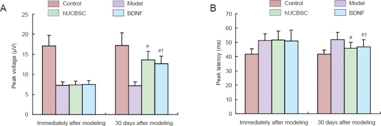
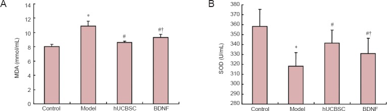
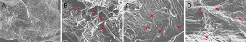
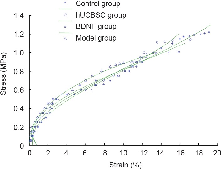
Treatment for optic nerve injury by brain-derived neurotrophic factor or the transplantation of human umbilical cord blood stem cells has gained progress, but analysis by biomechanical indicators is rare. Rabbit models of optic nerve injury were established by a clamp. At 7 days after injury, the vitreous body received a one-time injection of 50 μg brain-derived neurotrophic factor or 1 × 10(6) human umbilical cord blood stem cells. After 30 days, the maximum load, maximum stress, maximum strain, elastic limit load, elastic limit stress, and elastic limit strain had clearly improved in rabbit models of optical nerve injury after treatment with brain-derived neurotrophic factor or human umbilical cord blood stem cells. The damage to the ultrastructure of the optic nerve had also been reduced. These findings suggest that human umbilical cord blood stem cells and brain-derived neurotrophic factor effectively repair the injured optical nerve, improve biomechanical properties, and contribute to the recovery after injury.

Keywords: biomechanical properties; brain-derived neurotrophic factor; human umbilical cord blood stem cells; nerve regeneration; neural regeneration; optic nerve injury.


Conflict of interest statement

Conflicts of interest: None declared.

Figures

Figure 1
Figure 1
Effects of BDNF or hUCBSCs on electrophysiological properties of rabbit optic nerve. (A) Peak voltage (μV); (B) peak latency (ms). Data are expressed as the mean ± SD, with 16 rabbits in each group. Intergroup comparison was done using the Scheffe's methods. #P < 0.05, vs. model group; †P < 0.05, vs. hUCBSC group. BDNF: Brain-derived neurotrophic factor; hUCBSC(s): human umbilical cord blood stem cell(s).
Figure 2
Figure 2
Effects of BDNF or hUCBSCs on serum MDA content (A) and SOD activity (B) in rabbits with optic nerve injury. Data are expressed as the mean ± SD, with 16 rabbits in each group. Intergroup comparison was done using Scheffe's method. *P < 0.05, vs. control group; #P < 0.05, vs. model group; †P < 0.05, vs. hUCBSC group. BDNF: Brain-derived neurotrophic factor; hUCBSC(s): human umbilical cord blood stem cell(s); MDA: malondialdehyde; SOD: superoxide dismutase.
Figure 3
Figure 3
Effects of BDNF or hUCBSCs on ultrastructure of injured optic nerve (scanning electron microscope, × 100). (A) Normal morphology of optic nerve cells in rabbits of the control group; (B) evident injury to optic nerve of rabbits in the model group; some nerve fibers were ordered on the cross section in the hUCBSC group (C) and BDNF group (D). Arrows show nerve fibers. BDNF: Brain-derived neurotrophic factor; hUCBSC(s): human umbilical cord blood stem cell(s).
Figure 4
Figure 4
Effects of BDNF or hUCBSCs on stress-strain curves of injured optic nerve. BDNF: Brain-derived neurotrophic factor; hUCBSC(s): human umbilical cord blood stem cell(s).

Similar articles

Cited by

References

    1. Bar-Ilan A, Naveh N, Weissman C, Belkin M, Schwartz M. Prostaglandin E2 changes in the retina and optic nerve of an eye with injured optic nerve. Neuroscience. 1991;45:221–225. - PubMed
    1. Dagnelie G. Retinal implants: emergence of a multidisciplinary field. Curr Opin Neurol. 2012;25:67–75. - PMC - PubMed
    1. Hollander A, D’Onofrio PM, Magharious MM, Lysko MD, Koeberle PD. Quantitative retinal protein analysis after optic nerve transection reveals a neuroprotective role for hepatoma-derived growth factor on injured retinal ganglion cells. Invest Ophthalmol Vis Sci. 2012;53:3973–3989. - PubMed
    1. Jiang B, Zhang P, Zhou D, Zhang J, Xu X, Tang L. Intravitreal transplantation of human umbilical cord blood stem cells protects rats from traumatic optic neuropathy. PLoS One. 2013;8:e69938. - PMC - PubMed
    1. Jiang YY, Xu HT, Liu JX, Li P, Wu YZ. Biomechanical analysis of optic nerve injury treated by compound light granules and ciliary neurotrophic factor. Neural Regen Res. 2012;7:2889–2900. - PMC - PubMed

LinkOut - more resources