

Differential induction of PD-1/PD-L1 in Neuroimmune cells by drug of abuse
- PMID: 26330898
- PMCID: PMC4550211
Differential induction of PD-1/PD-L1 in Neuroimmune cells by drug of abuse
Abstract
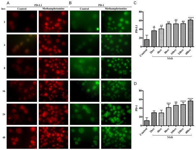
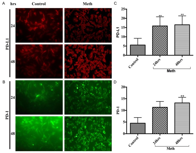
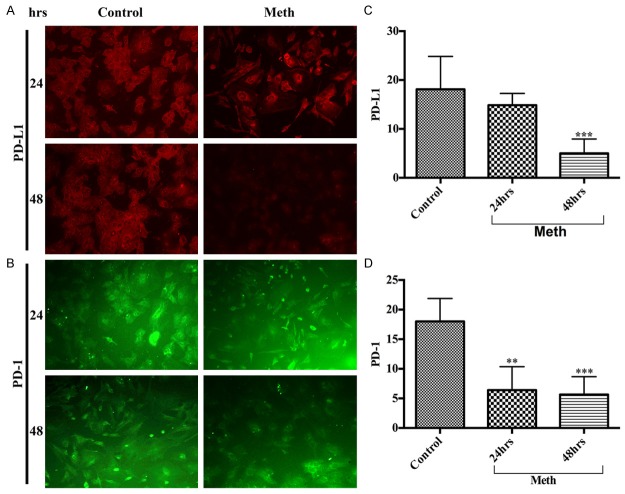
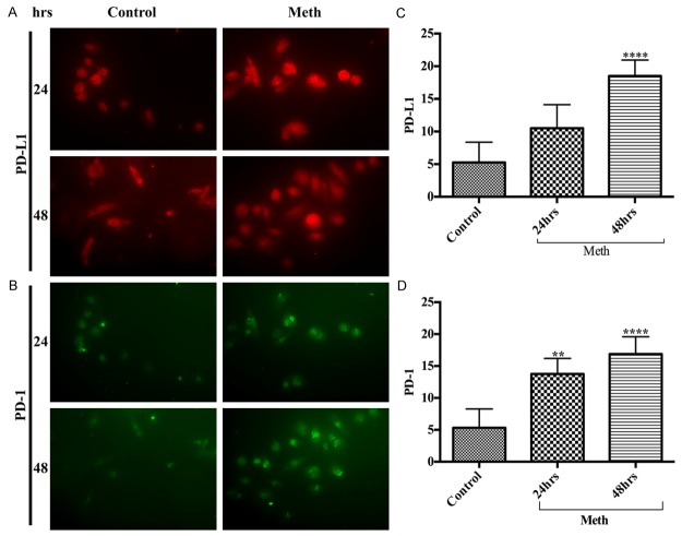
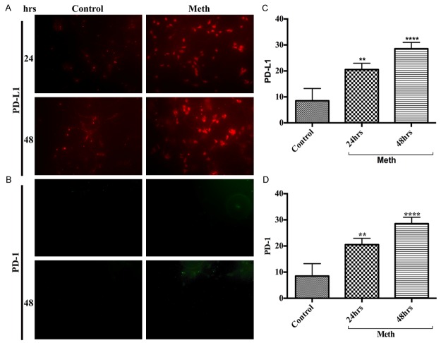
Interaction of programmed cell death protein 1 (PD-1) and programmed death-ligand 1 (PD-L1) plays a critical role in regulating the delicate balance between protective immunity and tolerance. Human neuroimmune cells express very low or undetectable levels of PD-1/PD-L1 in normal physiological condition.We seek to examine if exposure of these cells to drug of abuse such as methamphetamine (METH) alters the profile of PD-1/PD-L1 levels, thereby dampens the innate immune response of the host cells. Thus, we assessed the changes in the levels of PD-1/PD-L1 in primary human macrophages, brain endothelial cells (hBECs), astrocytes, microglia, and neurons after exposure to METH. We observed that stimulation of these neuroimmune cells by METH responded differentially to PD-1/PD-L1 expression. Stimulation of macrophage culture with 50 μM of METH exhibited immediate gradual upregulation of PD-L1, while increase in PD-1 took 2-4 hours later than PD-L1. The response of hBECs to PD-1/PD-L1 induction occurred at 24 hours, while increase of PD-1/PD-L1 levels in neurons and microglia was immediate following METH exposure. We found that astrocytes expressed moderate levels of endogenous PD-1/PD-L1, which was diminished by METH exposure. Our findings show a differential expression of PD-1/PD-L1 in neuroimmune cells in response to METH stimulation, suggesting that PD-1/PD-L1 interplay in these cell types could orchestrate the intercellular interactive communication for neuronal death or protection in the brain environment.
Keywords: Methamphetamine; neurodegeneration; neuroimmune cells; neuroinflammation; programmed cell death 1.
Figures
Similar articles
-
Alcohol induces programmed death receptor-1 and programmed death-ligand-1 differentially in neuroimmune cells.Alcohol. 2020 Aug;86:65-74. doi: 10.1016/j.alcohol.2020.03.009. Epub 2020 Mar 26. Alcohol. 2020. PMID: 32224220
-
Immune regulation of canine tumour and macrophage PD-L1 expression.Vet Comp Oncol. 2017 Jun;15(2):534-549. doi: 10.1111/vco.12197. Epub 2016 Feb 4. Vet Comp Oncol. 2017. PMID: 26842912
-
Enhanced Expression of PD-L1 on Microglia After Surgical Brain Injury Exerts Self-Protection from Inflammation and Promotes Neurological Repair.Neurochem Res. 2019 Nov;44(11):2470-2481. doi: 10.1007/s11064-019-02864-8. Epub 2019 Sep 3. Neurochem Res. 2019. PMID: 31482256
-
Glial Cell Expression of PD-L1.Int J Mol Sci. 2019 Apr 4;20(7):1677. doi: 10.3390/ijms20071677. Int J Mol Sci. 2019. PMID: 30987269 Free PMC article. Review.
-
HIV-1, methamphetamine and astrocytes at neuroinflammatory Crossroads.Front Microbiol. 2015 Oct 27;6:1143. doi: 10.3389/fmicb.2015.01143. eCollection 2015. Front Microbiol. 2015. PMID: 26579077 Free PMC article. Review.
Cited by
-
Neurological Immune-Related Adverse Events Induced by Immune Checkpoint Inhibitors.Biomedicines. 2024 Jun 13;12(6):1319. doi: 10.3390/biomedicines12061319. Biomedicines. 2024. PMID: 38927526 Free PMC article. Review.
-
Programmed Cell Death Protein 1 Blockade Reduces Glycogen Synthase Kinase 3β Activity and Tau Hyperphosphorylation in Alzheimer's Disease Mouse Models.Front Cell Dev Biol. 2021 Dec 16;9:769229. doi: 10.3389/fcell.2021.769229. eCollection 2021. Front Cell Dev Biol. 2021. PMID: 34977020 Free PMC article.
-
Inflammatory CNS disease caused by immune checkpoint inhibitors: status and perspectives.Nat Rev Neurol. 2017 Dec;13(12):755-763. doi: 10.1038/nrneurol.2017.144. Epub 2017 Nov 6. Nat Rev Neurol. 2017. PMID: 29104289 Review.
-
The immunomodulatory effect of microglia on ECM neuroinflammation via the PD-1/PD-L1 pathway.CNS Neurosci Ther. 2022 Jan;28(1):46-63. doi: 10.1111/cns.13760. Epub 2021 Nov 11. CNS Neurosci Ther. 2022. PMID: 34766463 Free PMC article.
References
-
- UNODOC. United Nations Office on Drugs and Crime. Vienna: 2011. World drug report.
-
- Grant KM, Kelley SS, Agrawal S, Meza JL, Meyer JR, Romberger DJ. Methamphetamine use in rural Midwesterners. Am J Addict. 2007;16:79–84. - PubMed
-
- Cadet JL, Jayanthi S, Deng X. Speed kills: cellular and molecular bases of methamphetamine-induced nerve terminal degeneration and neuronal apoptosis. FASEB J. 2003;17:1775–1788. - PubMed
-
- Davidson C, Gow AJ, Lee TH, Ellinwood EH. Methamphetamine neurotoxicity: necrotic and apoptotic mechanisms and relevance to human abuse and treatment. Brain Res Brain Res Rev. 2001;36:1–22. - PubMed
Grants and funding
LinkOut - more resources
Full Text Sources
Research Materials