Analysis of Clinical Feature and Management of Fish Bone Ingestion of Upper Gastrointestinal Tract
- PMID: 26330922
- PMCID: PMC4553358
- DOI: 10.3342/ceo.2015.8.3.261
Analysis of Clinical Feature and Management of Fish Bone Ingestion of Upper Gastrointestinal Tract
Abstract
Objectives: Fish bone impaction in the upper gastrointestinal tract is a common reason for patients to seek emergent care. The aim of this study was to find a clinical characteristics of patients with fish bone impaction in the upper gastrointestinal tract.
Methods: The study was conducted on 286 fish bone ingestion patients who complained of dysphagia and irritation after eating fish. The patients were treated according to the hospital protocol regarding the removal of fish bone. The parameters for the analysis included the age and sex of the patients, location and characteristics of the foreign body, method of removal, and type of fish.
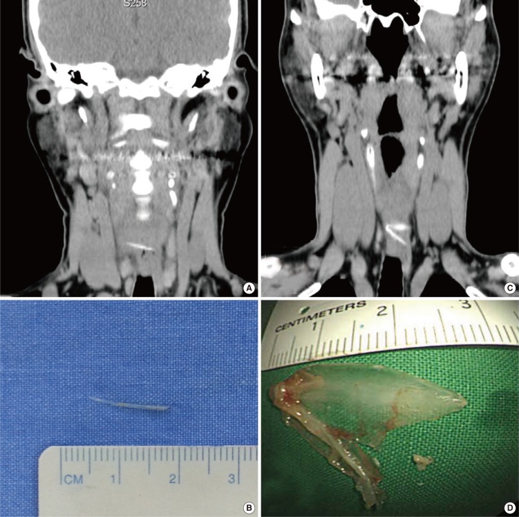
Results: The fish bone could be observed by the physical examination in the oral cavity and laryngopharynx in 198 patients (69.23%). For those patients in whom the foreign body could not be observed in oral cavity and laryngopharynx, noncontrast computed tomography (CT) (from nasopharynx to diaphragm) was performed. The fish bone was discovered in the esophagus of 66 patients (23.08%). The esophageal fish bone was successfully removed by transnasal flexible esophagoscopy (TNE) in 55 patients, the fish bone moved to the stomach in 10 patients and one fish bone was removed by rigid esophagoscopy due to esophageal abscess. The esophageal fish bone was mostly found in patients aged 50 years and older.
Conclusion: Fish bone foreign body ingestion in the esophagus appeared to be more common in older patients. Incorporating noncontrast CT and TNE can facilitate decision-making and adequate treatment for patients with fish bone impactions.
Keywords: Aged; Bone and Bones; Endoscopy; Fishes; Foreign Bodies.
Conflict of interest statement
Figures


References
-
- Vizcarrondo FJ, Brady PG, Nord HJ. Foreign bodies of the upper gastrointestinal tract. Gastrointest Endosc. 1983 Aug;29(3):208–210. - PubMed
-
- Henderson CT, Engel J, Schlesinger P. Foreign body ingestion: review and suggested guidelines for management. Endoscopy. 1987 Mar;19(2):68–71. - PubMed
-
- Nandi P, Ong GB. Foreign body in the oesophagus: review of 2394 cases. Br J Surg. 1978 Jan;65(1):5–9. - PubMed
-
- Webb WA. Management of foreign bodies of the upper gastrointestinal tract. Gastroenterology. 1988 Jan;94(1):204–216. - PubMed
-
- Devanesan J, Pisani A, Sharma P, Kazarian KK, Mersheimer WL. Metallic foreign bodies in the stomach. Arch Surg. 1977 May;112(5):664–665. - PubMed
LinkOut - more resources
Full Text Sources
Other Literature Sources