

Demineralized Bone Matrix Injection in Consolidation Phase Enhances Bone Regeneration in Distraction Osteogenesis via Endochondral Bone Formation
- PMID: 26330963
- PMCID: PMC4553289
- DOI: 10.4055/cios.2015.7.3.383
Demineralized Bone Matrix Injection in Consolidation Phase Enhances Bone Regeneration in Distraction Osteogenesis via Endochondral Bone Formation
Abstract
Background: Distraction osteogenesis (DO) is a promising tool for bone and tissue regeneration. However, prolonged healing time remains a major problem. Various materials including cells, cytokines, and growth factors have been used in an attempt to enhance bone formation. We examined the effect of percutaneous injection of demineralized bone matrix (DBM) during the consolidation phase on bone regeneration after distraction.
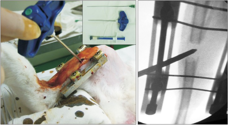
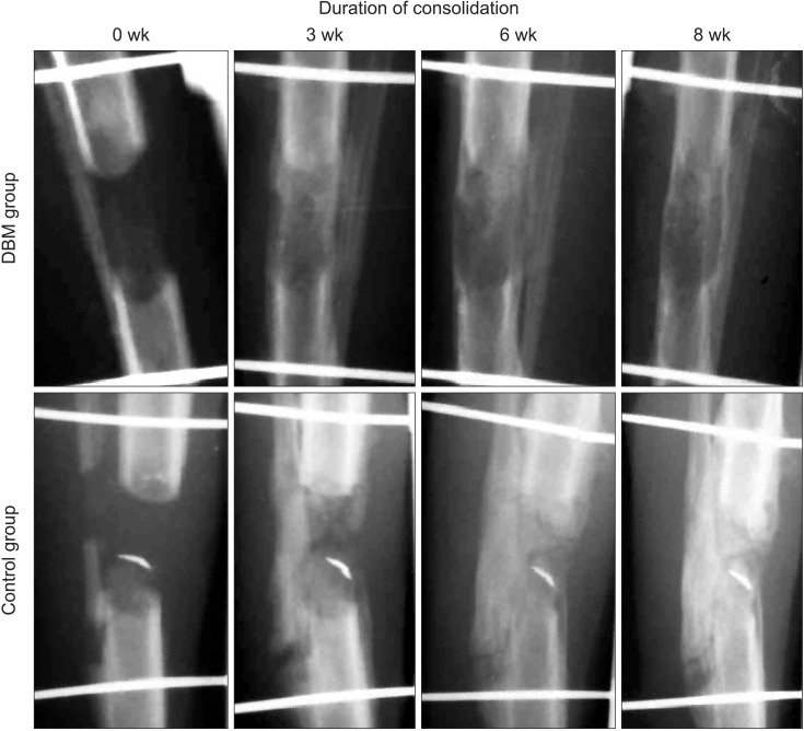
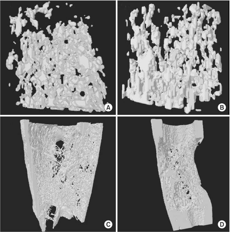
Methods: The immature rabbit tibial DO model (20 mm length-gain) was used. Twenty-eight animals received DBM 100 mg percutaneously at the end of distraction. Another 22 animals were left without further procedure (control). Plain radiographs were taken every week. Postmortem bone dual-energy X-ray absorptiometry and micro-computed tomography (micro-CT) studies were performed at the third and sixth weeks of the consolidation period and histological analysis was performed.
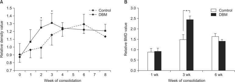
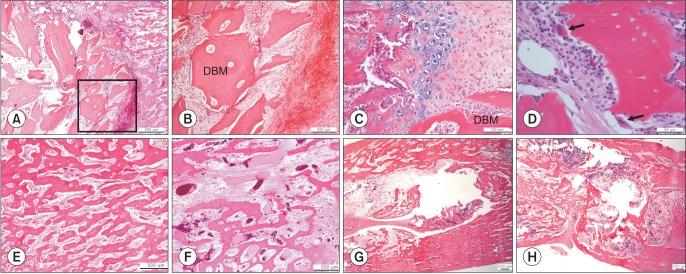
Results: The regenerate bone mineral density was higher in the DBM group when compared with that in the saline injection control group at the third week postdistraction. Quantitative analysis using micro-CT revealed larger trabecular bone volume, higher trabecular number, and less trabecular separation in the DBM group than in the saline injection control group. Cross-sectional area and cortical thickness at the sixth week postdistraction, assessed using micro-CT, were greater in the regenerates of the DBM group compared with the control group. Histological evaluation revealed higher trabecular bone volume and trabecular number in the regenerate of the DBM group. New bone formation was apparently enhanced, via endochondral ossification, at the site and in the vicinity of the injected DBM. DBM was absorbed slowly, but it remained until the sixth postoperative week after injection.
Conclusions: DBM administration into the distraction gap at the end of the distraction period resulted in a significantly greater regenerate bone area, trabecular number, and cortical thickness in the rabbit tibial DO model. These data suggest that percutaneous DBM administration at the end of the distraction period or in the early consolidation period may stimulate regenerate bone formation and consolidation in a clinical situation with delayed bone healing during DO.
Keywords: Bone development; Bone substitute; Distraction; Osteogenesis.
Conflict of interest statement
Figures
Similar articles
-
Percutaneous CO2 Treatment Accelerates Bone Generation During Distraction Osteogenesis in Rabbits.Clin Orthop Relat Res. 2020 Aug;478(8):1922-1935. doi: 10.1097/CORR.0000000000001288. Clin Orthop Relat Res. 2020. PMID: 32732577 Free PMC article.
-
[Experimental study on promoting bone consolidation by using platelet-rich plasma and decalcified bone matrix during distraction osteogenesis].Zhongguo Xiu Fu Chong Jian Wai Ke Za Zhi. 2011 Jun;25(6):661-7. Zhongguo Xiu Fu Chong Jian Wai Ke Za Zhi. 2011. PMID: 21735776 Chinese.
-
[Effect of "accordion" technique on bone consolidation during distraction osteogenesis].Zhongguo Xiu Fu Chong Jian Wai Ke Za Zhi. 2018 May 15;32(5):558-567. doi: 10.7507/1002-1892.201712094. Zhongguo Xiu Fu Chong Jian Wai Ke Za Zhi. 2018. PMID: 29806343 Free PMC article. Chinese.
-
Management of Osseous Defects in the Tibia: Utilization of External Fixation, Distraction Osteogenesis, and Bone Transport.Clin Podiatr Med Surg. 2021 Jan;38(1):111-116. doi: 10.1016/j.cpm.2020.09.006. Clin Podiatr Med Surg. 2021. PMID: 33220740 Review.
-
Strategies of enhancing bone regenerate formation in distraction osteogenesis.Connect Tissue Res. 2018 Jan;59(1):1-11. doi: 10.1080/03008207.2017.1288725. Epub 2017 Mar 8. Connect Tissue Res. 2018. PMID: 28165797 Review.
Cited by
-
Expert consensus on the bone repair strategy for osteoporotic fractures in China.Front Endocrinol (Lausanne). 2022 Oct 26;13:989648. doi: 10.3389/fendo.2022.989648. eCollection 2022. Front Endocrinol (Lausanne). 2022. PMID: 36387842 Free PMC article. Review.
-
An Inexpensive 3D Printed Mouse Model of Successful, Complication-free Long Bone Distraction Osteogenesis.Plast Reconstr Surg Glob Open. 2023 Feb 13;11(2):e4674. doi: 10.1097/GOX.0000000000004674. eCollection 2023 Feb. Plast Reconstr Surg Glob Open. 2023. PMID: 36798717 Free PMC article.
-
Use of allograft bone matrix in clinical orthopedics.Regen Med. 2024 May 3;19(5):247-256. doi: 10.1080/17460751.2024.2353473. Epub 2024 Jul 19. Regen Med. 2024. PMID: 39028538 Free PMC article. Review.
-
The efficacy of rhBMP-2 loaded hydrogel composite on bone formation around dental implants in mandible bone defects of minipigs.Biomater Res. 2020 Feb 3;24:5. doi: 10.1186/s40824-020-0183-9. eCollection 2020. Biomater Res. 2020. PMID: 32042440 Free PMC article.
-
Percutaneous CO2 Treatment Accelerates Bone Generation During Distraction Osteogenesis in Rabbits.Clin Orthop Relat Res. 2020 Aug;478(8):1922-1935. doi: 10.1097/CORR.0000000000001288. Clin Orthop Relat Res. 2020. PMID: 32732577 Free PMC article.
References
-
- Catagni MA, Guerreschi F, Holman JA, Cattaneo R. Distraction osteogenesis in the treatment of stiff hypertrophic nonunions using the Ilizarov apparatus. Clin Orthop Relat Res. 1994;(301):159–163. - PubMed
-
- Paley D. Treatment of tibial nonunion and bone loss with the Ilizarov technique. Instr Course Lect. 1990;39:185–197. - PubMed
-
- Moseley CF. Leg lengthening: a review of 30 years. Clin Orthop Relat Res. 1989;(247):38–43. - PubMed
-
- Korzinek K, Tepic S, Perren SM. Limb lengthening and three-dimensional deformity corrections: a retrospective clinical study. Arch Orthop Trauma Surg. 1990;109(6):334–340. - PubMed
Publication types
MeSH terms
Substances
LinkOut - more resources
Full Text Sources
Other Literature Sources
Miscellaneous
