

doi: 10.1038/bonekey.2015.101.
eCollection 2015.
Assessment of muscle mass and strength in mice
Affiliations
- PMID: 26331011
- PMCID: PMC4549925
- DOI: 10.1038/bonekey.2015.101
Item in Clipboard
Assessment of muscle mass and strength in mice
Bonekey Rep.
.
Abstract
Muscle weakness is an important phenotype of many diseases that is linked to impaired locomotion and increased mortality. The force that a muscle can generate is determined predominantly by muscle size, fiber type and the excitation-contraction coupling process. Here we describe methods for the histological assessment of whole muscle to determine fiber cross-sectional area and fiber type, determination of changes in myocyte size using C2C12 cells, in vivo functional tests and measurement of contractility in dissected whole muscles. The extensor digitorum longus and soleus muscles are ideally suited for whole-muscle contractility, and dissection of these muscles is described.
Conflict of interest statement
The authors declare no conflict of interest.
Figures
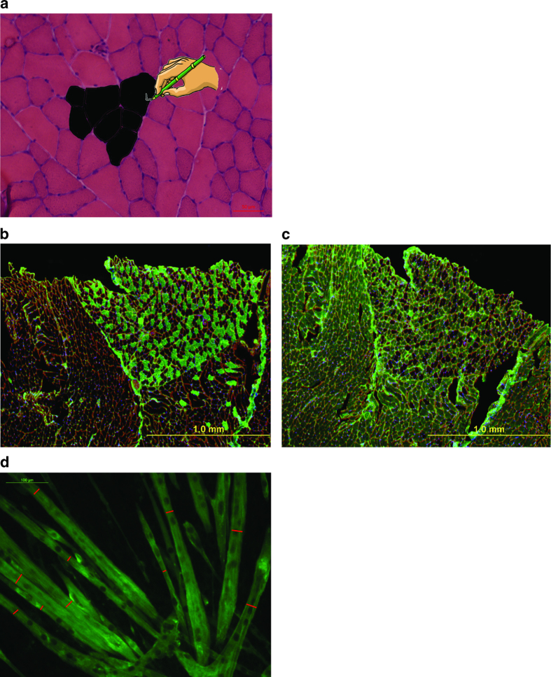
Histological analysis of skeletal muscle. (a) Photomicrograph of muscle cross-section stained by H&E with a schematic on how to use the ImgaeJ software to outline and measure the cross-sectional area of the fibers. Magnification 20X, scale bar: 50 μm. (b–c) Representative fiber typing. Consecutive 8-μm thick sections from liquid nitrogen-cooled isopentane frozen gastrocnemius muscle were stained for (b) Myosin Heavy Chain slow (BA-D5 mouse antibody diluted 1:50; green) or (c) Myosin Heavy Chain fast (SC-71 mouse antibody diluted 1:50; green) and visualized by anti-mouse-IgG Alexa Fluor-488 (Life Technologies) secondary antibody diluted 1:1000 in PBS. (d) Representative photomicrograph of C2C12 murine myotube cultures fixed in ice-cold 4% paraformaldehyde in PBS and subsequently incubated in anti-myosin heavy chain (MF-20 clone, Developmental Studies Hybridoma Bank) monoclonal antibody diluted 1:1000 in PBS overnight at 4 °C. Myotubes were then visualized using anti-mouse-IgG Alexa Fluor-488 (Life Technologies) secondary antibody diluted 1:1000 in PBS at room temperature for 30–45 min. Red bars indicate the minimum diameter for each fiber.
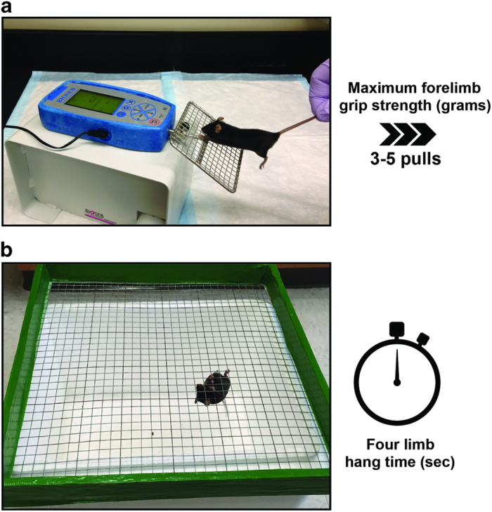
In vivo assessment of muscle strength. (a) Forelimb grip strength test of mouse grasping wire screen as it is being pulled by the tail horizontally relative to the force meter. The mouse is pulled steadily until it loses grip. The peak force is measured by the force meter. (b) Four limb-hanging test (Kondziella's inverted screen test) of mouse grasping wire screen as it is inverted. The time of sustained limb tension to oppose their weight is measured (hang time and minimal holding impulse).
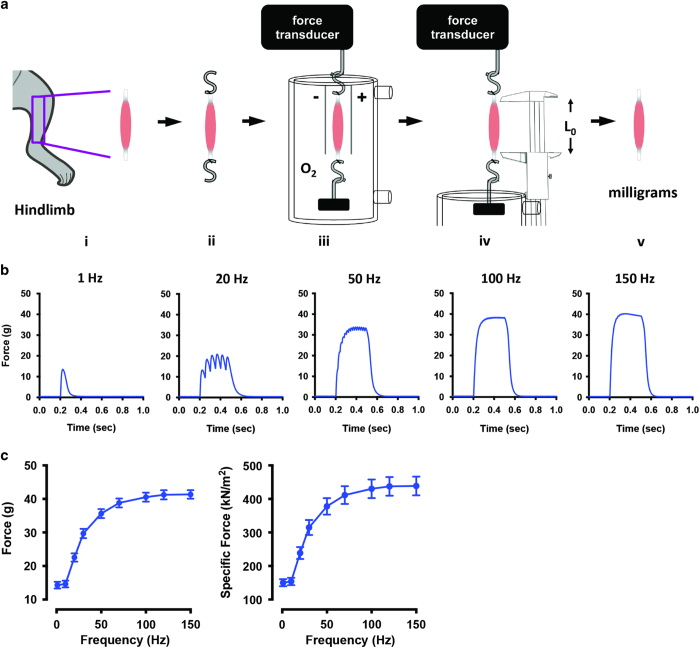
Isolated whole muscle contractility of mouse skeletal muscle. (a) Schematic illustration of the steps to determine isolated whole muscle contractility of skeletal muscle: (i), the muscle is excised from the lower hind limb; (ii), stainless steel hooks are attached via silk suture to the tendons; (iii), the muscle is bathed in physiological solution (Tyrode's buffer) bubbled with oxygen and stimulated to contract via platinum electrodes, and force is recorded by the force transducer; (iv), the optimal length (L0) is measured using vernier style calipers; (v), the semi-dry weight of the muscle is measured using an analytical balance (mg). (b) Representative single muscle data at various frequencies using the EDL excised from 28-week-old C57BL/6 mice. (c) Representative absolute force and specific force of the EDL from 28-week-old C57BL/6 mice (n=10±s.e.m.).
References
-
- Bonetto A, Penna F, Minero VG, Reffo P, Bonelli G, Baccino FM et al.. Deacetylase inhibitors modulate the myostatin/follistatin axis without improving cachexia in tumor-bearing mice. Curr Cancer Drug Targets 2009; 9: 608–616. - PubMed
Grants and funding
LinkOut - more resources
Full Text Sources
Other Literature Sources