Review
. 2014 Jun 13:18:5.
doi: 10.1186/2055-7124-18-5. eCollection 2014.

Nanofibrous scaffolds in biomedical applications

Affiliations
Review

Nanofibrous scaffolds in biomedical applications

Kailash Chandra Gupta et al. Biomater Res. .

Abstract

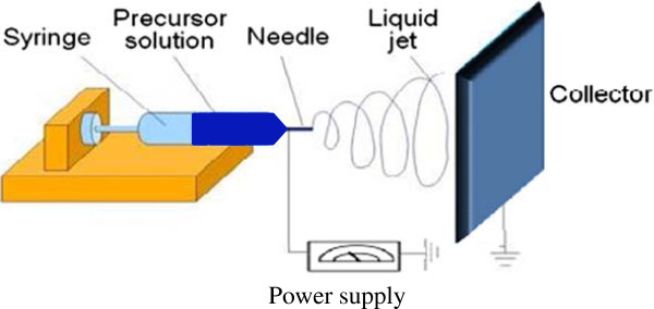
Nanofibrous scaffolds are artificial extracellular matrices which provide natural environment for tissue formation. In comparison to other forms of scaffolds, the nanofibrous scaffolds promote cell adhesion, proliferation and differentiation more efficiently due to having high surface to volume ratio. Although scaffolds for tissue engineering have been fabricated by various techniques but electrospun nanofibrous scaffolds have shown great potential in the fields of tissue engineering and regeneration. This review highlights the applications and importance of electrospun nanofibrous scaffolds in various fields of biomedical applications ranging from drug delivery to wound healing. Attempts have also been made to highlights the advantages and disadvantages of nanofirbous scaffolds fabricated for biomedical applications using technique of electrospinning. The role of various factors controlling drug distribution in electrospun nanofibrous scaffolds is also discussed to increase the therapeutic efficiency of nanofibrous scaffolds in wound healing and drug delivery applications.

Keywords: Bioactive agent; Drug delivery; Electrospinning; Tissue engineering; Wound healing.


Figures

Figure 1
Figure 1
An electrospinning setup showing the arrangement for syringe, precursor solution, needle, liquid jet, collector, and power supply.
Figure 2
Figure 2
SEM micrographs of electrospun nanofibrous scaffolds using a PHBV/gelatin solution at TFE 6 wt%; (a) 30/70, (b) 50/50, and (c) 70/30 (adapted from reference [127] ).
Figure 3
Figure 3
SEM micrographs of electrospun nanofibrous scaffolds using TFE solutions with different amount of PHBV/gelatin (50/50); (a) 2 wt%, (b) 4 wt%, (c) 6 wt%, and (d) 8 wt% (adapted from reference [127] ).
Figure 4
Figure 4
AFM phase images of nanofibrous scaffold surface; (a) PHBV and (b) PHBV–Col (Adapted from reference [128] ).

References

    1. Rošic R, Kocbek P, Pelipenko J, Kristl J, Baumgartner S. Nanofibers and their biomedical use. Acta Pharma. 2013;63:295. doi: 10.2478/acph-2013-0024. - DOI - PubMed
    1. Haider A, Gupta KC, Kang IK. BioMed Res Int. 2014. Morphological effects of HA on the cell compatibility of electrospun HA/PLGA composite nanofiber scaffolds. - PMC - PubMed
    1. Li X, Kanjwal MA, Lin L, Chronakis IS. Electrospun polyvinyl-alcohol nanofibers as oral fast-dissolving delivery system of caffeine and riboflavin. Colloids Surf B Biointerf. 2013;103:1828. doi: 10.1016/j.colsurfb.2012.10.016. - DOI - PubMed
    1. Illangakoon UE, Nazir T, Williams GR, Chatterton NP. Mebeverine-loaded electrospun nanofibers: physicochemical characterization and dissolution studies. J Pharma Sci. 2014;103:283. doi: 10.1002/jps.23759. - DOI - PubMed
    1. Karthikeyan K, Guhathakarta S, Rajaram R, Korrapati PS. Electrospun zein/eudragit nanofibers based dual drug delivery system for the simultaneous delivery of aceclofenac and pantoprazole. Int J Pharm. 2012;438:17. doi: 10.1016/j.ijpharm.2012.07.075. - DOI - PubMed

LinkOut - more resources