

Electron beam effect on biomaterials I: focusing on bone graft materials
- PMID: 26331080
- PMCID: PMC4552193
- DOI: 10.1186/s40824-015-0031-5
Electron beam effect on biomaterials I: focusing on bone graft materials
Abstract
Background: To develop biocompatible bony regeneration materials, allogenic, xenogenic and synthetic bones have been irradiated by an electron beam to change the basic structures of their inorganic materials. The optimal electron beam energy and individual dose have not been established for maximizing the bony regeneration capacity in electron beam irradiated bone.
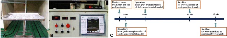
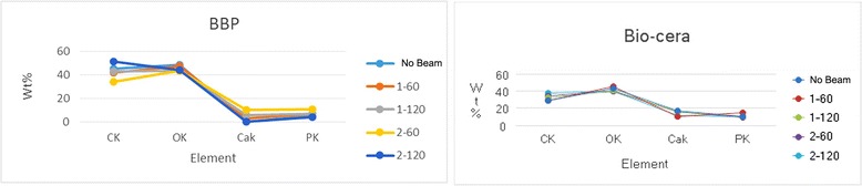
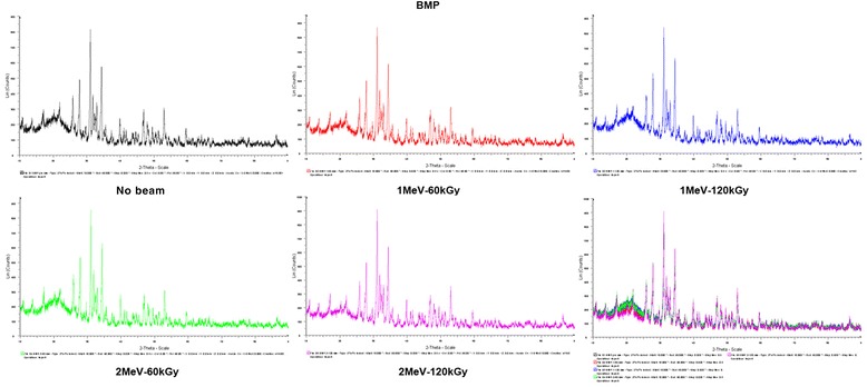
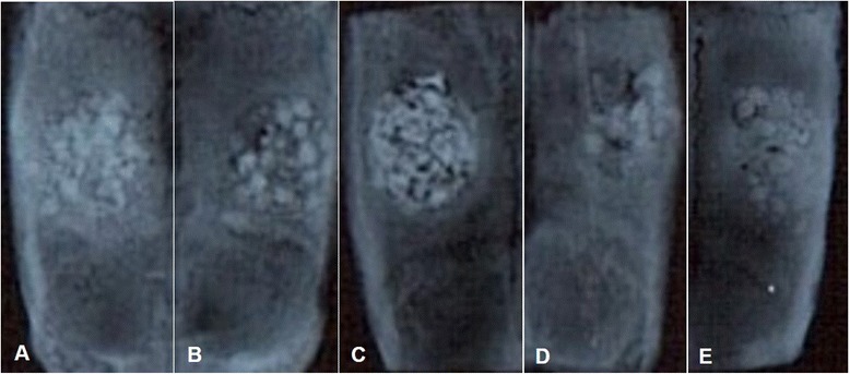
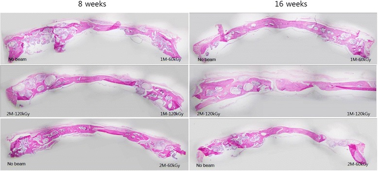
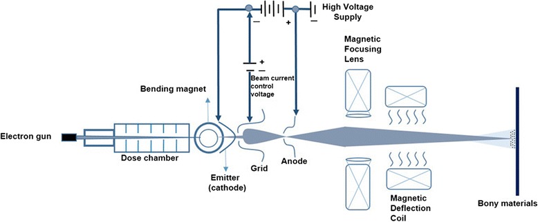
Results: Commercial products consisting of four allogenic bones, six xenogenic bones, and six synthetic bones were used in this study. We used 1.0-MeV and 2.0 MeV linear accelerators (power: 100 KW, pressure; 115 kPa, temperature; -30 to 120°C, sensor sensitivity: 0.1-1.2 mV/kPa, generating power sensitivity: 44.75 mV/kPa, supply voltage: 50.25 V), and a microtrone with different individual irradiation doses such as 60 kGy and 120 kGy. Additional in vitro analyses were performed by elementary analysis using field emission scanning electron microscopy (FE-SEM), scanning electron microscopy (SEM), X-ray diffraction (XRD) and confocal laser scanning microscopy (CLSM). In vivo clinical, radiographic, and micro-computed tomography (Micro-CT) with bone marrow density (BMD) analysis was performed in 8- and 16-week-old Spraque-Dawley rats with calvarial defect grafts.
Conclusions: Electron beam irradiation of bony substitutes has four main effects: the cross-linking of biphasic calcium phosphate bony apatite, chain-scissioning, the induction of rheological changes, and microbiological sterilization. These novel results and conclusions are the effects of electron beam irradiation.
Keywords: Allogenic bone; Bony regeneration; Electron beam irradiation; Synthetic bone; Xenogenic bone.
Figures


References
-
- Kim SM, Eo MY, Kang JY, Myoung H, Choi EK, Lee SK, et al. Bony regeneration effect of electron-beam irradiated hydroxyapatite and tricalcium phosphate mixtures with 7 to 3 ratio in the calvarial defect model of rat. Tissue Engineering Regenerative Medicine. 2013;9:24–32.
-
- Park JM, Kim SM, Kim MK, Park YW, Myoung H, Lee BC, et al. Effect of electron-beam irradiation on the artificial bone substitutes composed of hydroxyapatite and tricalcium phosphate mixtures with type I collagen. J Korean Assoc Maxillofac Plast Reconstr Surg. 2013;35:38–50.
-
- Laurell B, Föll E. Electron-beam accelerators for new applications. RadTech Europe 2011 Exhibition & Conference for Radiation Curing. Electron Crosslinking AB; 2011
-
- Kim JH, Kim SM, Kim JH, Kwon KJ, Park YW. Effect of type I collagen on hydroxyapatite and tricalcium phosphate mixtures in rat calvarial bony defects. J Kor Oral Maxillofac Surg. 2008;34:36–48.
-
- Clark G: Staining procedure, 4th edn. Wiliams & Wilkins, Baltimore.
LinkOut - more resources
Full Text Sources
Other Literature Sources
