. 2015 Aug;5(4):323-9.
doi: 10.3978/j.issn.2223-3652.2015.04.05.

Stent mal-apposition with resorption of intramural hematoma with spontaneous coronary artery dissection

Affiliations

Stent mal-apposition with resorption of intramural hematoma with spontaneous coronary artery dissection

Mathieu Lempereur et al. Cardiovasc Diagn Ther. 2015 Aug.

Abstract

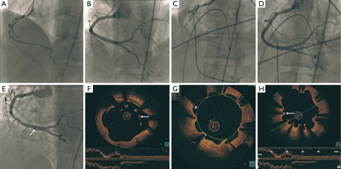
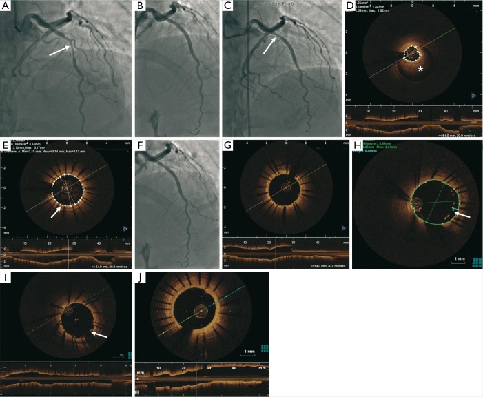
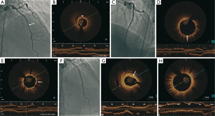
Spontaneous coronary artery dissection (SCAD) is an infrequent cause of acute coronary syndrome (ACS). Conservative management is typically recommended but revascularization may be necessary if ongoing ischemia or adverse anatomical characteristics are present. Percutaneous coronary intervention (PCI) of SCAD can be fraught with challenges, and intracoronary imaging with optical coherence tomography (OCT) may provide insights on optimizing the acute results and identify long-term stent-related adverse events. We report three cases of SCAD treated with drug-eluting stents (DES) with OCT follow-up showing stent mal-apposition at different stages of follow-up. The clinical significance of these OCT findings and management options are discussed.

Keywords: Angiography; arterial occlusive disease; coronary artery disease; optical coherence tomography (OCT); percutaneous coronary intervention (PCI).


Conflict of interest statement

Conflicts of Interest: J Saw has received unrestricted research grant supports (from the Canadian Institutes of Health Research, University of British Columbia Division of Cardiology, AstraZeneca, Abbott Vascular, St Jude Medical, Boston Scientific, and Servier), speaker honoraria (AstraZeneca, St Jude Medical, Boston Scientific, Bayer and Sunovion), consultancy and advisory board honoraria (AstraZeneca, St Jude Medical, Boston Scientific, and Abbott Vascular), and proctorship honoraria (St Jude Medical and Boston Scientific). A Fung has received proctorship honoraria from St Jude Medical.

Figures

Figure 1
Figure 1
Diffuse dissection of the proximal to distal RCA (A); stenting of the RCA with six DES (B); very late stent thrombosis of the RCA (C) treated with balloon angioplasty 40 months later (D); patent RCA stents, but diffuse peri-stent staining (arrow) 32 months later (E); OCT showing large arcs of malapposition (arrow) (F); areas of evagination (*) reaching 2.5 mm2 (G); and multiple areas of uncovered stent struts (arrow) (H). RCA, right coronary artery; DES, drug-eluting stents; OCT, optical coherence tomography.
Figure 2
Figure 2
OCT showing large arcs of malapposition, areas of evagination, and multiple areas of uncovered stent struts (1). OCT, optical coherence tomography. Available online: http://www.asvide.com/articles/619
Figure 3
Figure 3
Tubular 80% stenosis (arrow) in the mid LAD (A); PCI with a 3.0 mm × 24 mm DES (B); patent LAD stent but severe luminal narrowing proximal to the stent (arrow) 9 days later (C); OCT demonstrating IMH (*) proximal to the stent (D) and tacked-up IMH in the wall of the stented segment (arrow) (E); proximal IMH treated with 3.0 mm × 15 mm DES (F) with good stent expansion and apposition (G); OCT 2 days later revealed malapposition of the proximal LAD stent at the prior site of the IMH with gaps between the struts and the intima reaching 460 µm (arrow) (H); final results on OCT with residual gap of 270 µm (arrow) after balloon angioplasty (I); repeat OCT 30 months later showed complete endothelialization of the stent struts (J). LAD, left anterior descending; PCI, percutaneous coronary intervention; OCT, optical coherence tomography; IMH, intramural hematoma; DES, drug-eluting stents.
Figure 4
Figure 4
OCT demonstrating IMH proximal to the stent and tacked-up IMH in the wall of the stented segment (2). OCT, optical coherence tomography; IMH, intramural hematoma. Available online: http://www.asvide.com/articles/620
Figure 5
Figure 5
A total of 50% lesion in the proximal LAD and 70% tubular lesion in the mid LAD (arrow) (A); LAD dissection with IMH (*) showed on OCT (B); results after PCI with two DES (C); OCT 15 days later showed patent stents but mild malapposition at the proximal edge of the proximal stent (250 µm) (arrow) (D) and persistent IMH covered by the stents (arrow) (E); 2 years later, 50% in-stent restenosis in the distal segment of the stent (F) with a minimal lumen area of 2.6 mm2 (G) and complete endothelialization of the stent struts (H). LAD, left anterior descending; IMH, intramural hematoma; OCT, optical coherence tomography; PCI, percutaneous coronary intervention; DES, drug-eluting stents.
Figure 6
Figure 6
OCT 15 days later showed patent stents but mild malapposition at the proximal edge of the proximal stent (250 µm) and persistent IMH covered by the stents (3). OCT, optical coherence tomography; IMH, intramural hematoma. Available online: http://www.asvide.com/articles/621

References

    1. Lempereur M, Fung A, Saw J. OCT showing large arcs of malapposition, areas of evagination, and multiple areas of uncovered stent struts. Asvide 2015;2:072. Available online: http://www.asvide.com/articles/619
    1. Lempereur M, Fung A, Saw J. OCT demonstrating IMH proximal to the stent and tacked-up IMH in the wall of the stented segment. Asvide 2015;2:073. Available online: http://www.asvide.com/articles/620
    1. Lempereur M, Fung A, Saw J. OCT 15 days later showed patent stents but mild malapposition at the proximal edge of the proximal stent (250 µm) and persistent IMH covered by the stents. Asvide 2015;2:074. Available online: http://www.asvide.com/articles/621
    1. Saw J, Sedlak T, Ganesh SK, et al. Cardiology patient page. Spontaneous coronary artery dissection (SCAD). Circulation 2015;131:e3-5. - PubMed
    1. Saw J. Spontaneous coronary artery dissection. Can J Cardiol 2013;29:1027-33. - PubMed