

Molecular evidence that invasive adenocarcinoma can mimic prostatic intraepithelial neoplasia (PIN) and intraductal carcinoma through retrograde glandular colonization
- PMID: 26331372
- PMCID: PMC4715537
- DOI: 10.1002/path.4628
Molecular evidence that invasive adenocarcinoma can mimic prostatic intraepithelial neoplasia (PIN) and intraductal carcinoma through retrograde glandular colonization
Abstract
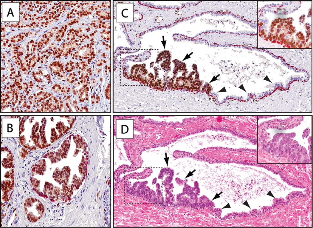
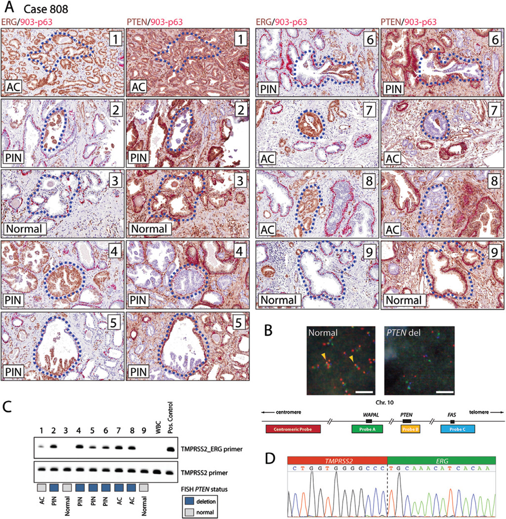
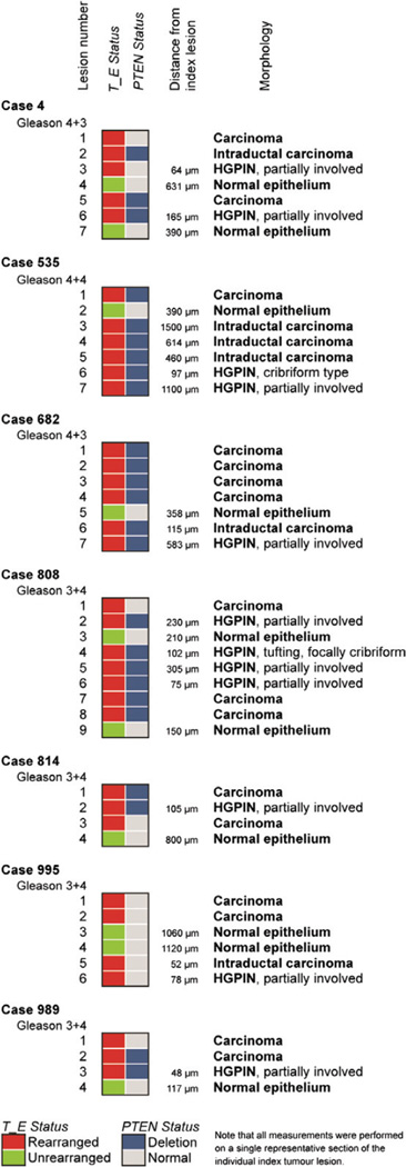
Prostate cancer often manifests as morphologically distinct tumour foci and is frequently found adjacent to presumed precursor lesions such as high-grade prostatic intraepithelial neoplasia (HGPIN). While there is some evidence to suggest that these lesions can be related and exist on a pathological and morphological continuum, the precise clonal and temporal relationships between precursor lesions and invasive cancers within individual tumours remain undefined. Here, we used molecular genetic, cytogenetic, and histological analyses to delineate clonal, temporal, and spatial relationships between HGPIN and cancer lesions with distinct morphological and molecular features. First, while confirming the previous finding that a substantial fraction of HGPIN lesions associated with ERG-positive cancers share rearrangements and overexpression of ERG, we found that a significant subset of such HGPIN glands exhibit only partial positivity for ERG. This suggests that such ERG-positive HGPIN cells either rapidly invade to form adenocarcinoma or represent cancer cells that have partially invaded the ductal and acinar space in a retrograde manner. To clarify these possibilities, we used ERG expression status and TMPRSS2-ERG genomic breakpoints as markers of clonality, and PTEN deletion status to track temporal evolution of clonally related lesions. We confirmed that morphologically distinct HGPIN and nearby invasive cancer lesions are clonally related. Further, we found that a significant fraction of ERG-positive, PTEN-negative HGPIN and intraductal carcinoma (IDC-P) lesions are most likely clonally derived from adjacent PTEN-negative adenocarcinomas, indicating that such PTEN-negative HGPIN and IDC-P lesions arise from, rather than give rise to, the nearby invasive adenocarcinoma. These data suggest that invasive adenocarcinoma can morphologically mimic HGPIN through retrograde colonization of benign glands with cancer cells. Similar clonal relationships were also seen for intraductal carcinoma adjacent to invasive adenocarcinoma. These findings represent a potentially undervalued indicator of pre-existing invasive prostate cancer and have significant implications for prostate cancer diagnosis and risk stratification.
Keywords: ERG; PTEN; clonality; ductal spreading; prostate cancer; prostatic intraepithelial neoplasia.
Copyright © 2015 Pathological Society of Great Britain and Ireland. Published by John Wiley & Sons, Ltd.
Figures

Similar articles
-
ERG and PTEN status of isolated high-grade PIN occurring in cystoprostatectomy specimens without invasive prostatic adenocarcinoma.Hum Pathol. 2016 Sep;55:117-25. doi: 10.1016/j.humpath.2016.04.017. Epub 2016 May 14. Hum Pathol. 2016. PMID: 27189342 Free PMC article.
-
ETS gene aberrations in atypical cribriform lesions of the prostate: Implications for the distinction between intraductal carcinoma of the prostate and cribriform high-grade prostatic intraepithelial neoplasia.Am J Surg Pathol. 2010 Apr;34(4):478-85. doi: 10.1097/PAS.0b013e3181d6827b. Am J Surg Pathol. 2010. PMID: 20220513
-
High-grade prostatic intraepithelial neoplasia, PIN-like carcinoma, ductal carcinoma, and intraductal carcinoma of the prostate.Mod Pathol. 2018 Jan;31(S1):S71-79. doi: 10.1038/modpathol.2017.138. Mod Pathol. 2018. PMID: 29297491 Review.
-
PTEN genomic deletion is an early event associated with ERG gene rearrangements in prostate cancer.BJU Int. 2011 Feb;107(3):477-85. doi: 10.1111/j.1464-410X.2010.09470.x. BJU Int. 2011. PMID: 20590547
-
Molecular Pathology of High-Grade Prostatic Intraepithelial Neoplasia: Challenges and Opportunities.Cold Spring Harb Perspect Med. 2019 Apr 1;9(4):a030403. doi: 10.1101/cshperspect.a030403. Cold Spring Harb Perspect Med. 2019. PMID: 30082453 Free PMC article. Review.
Cited by
-
Analysis of separate training and validation radical prostatectomy cohorts identifies 0.25 mm diameter as an optimal definition for "large" cribriform prostatic adenocarcinoma.Mod Pathol. 2022 Aug;35(8):1092-1100. doi: 10.1038/s41379-022-01009-7. Epub 2022 Feb 10. Mod Pathol. 2022. PMID: 35145197 Free PMC article.
-
Genetic Analysis of Intraductal Carcinoma of the Prostate Detected in High-Grade Prostatic Intraepithelial Neoplasia Cases.Cureus. 2024 Dec 21;16(12):e76165. doi: 10.7759/cureus.76165. eCollection 2024 Dec. Cureus. 2024. PMID: 39840193 Free PMC article.
-
MYC-driven increases in mitochondrial DNA copy number occur early and persist throughout prostatic cancer progression.JCI Insight. 2023 Dec 22;8(24):e169868. doi: 10.1172/jci.insight.169868. JCI Insight. 2023. PMID: 37971875 Free PMC article.
-
On cribriform prostate cancer.Transl Androl Urol. 2018 Feb;7(1):145-154. doi: 10.21037/tau.2017.12.33. Transl Androl Urol. 2018. PMID: 29594028 Free PMC article. Review.
-
Identification of intraductal-to-invasive spatial transitions in prostate cancer: proposal for a new unifying model on intraductal carcinogenesis.Histopathology. 2025 Jun;86(7):1091-1100. doi: 10.1111/his.15414. Epub 2025 Jan 30. Histopathology. 2025. PMID: 39888049 Free PMC article.
References
-
- Lindberg J, Klevebring D, Liu W, et al. Exome sequencing of prostate cancer supports the hypothesis of independent tumour origins. Eur Urol. 2013;63:347–353. - PubMed
-
- Bostwick DG, Shan A, Qian J. Matched foci of prostate carcinoma. Cancer. 1998;83:1995–2002. - PubMed
-
- Clark J, Attard G, Jhavar S. Prostate. Oncogene. 2007;27:1993–2003. - PubMed
Publication types
MeSH terms
Substances
Grants and funding
LinkOut - more resources
Full Text Sources
Other Literature Sources
Medical
Research Materials
