. 2015 Sep 2;10(9):e0137043.
doi: 10.1371/journal.pone.0137043. eCollection 2015.

Everolimus Stabilizes Podocyte Microtubules via Enhancing TUBB2B and DCDC2 Expression

Affiliations

Everolimus Stabilizes Podocyte Microtubules via Enhancing TUBB2B and DCDC2 Expression

Stefanie Jeruschke et al. PLoS One. .

Abstract

Background: Glomerular podocytes are highly differentiated cells that are key components of the kidney filtration units. The podocyte cytoskeleton builds the basis for the dynamic podocyte cytoarchitecture and plays a central role for proper podocyte function. Recent studies implicate that immunosuppressive agents including the mTOR-inhibitor everolimus have a protective role directly on the stability of the podocyte actin cytoskeleton. In contrast, a potential stabilization of microtubules by everolimus has not been studied so far.

Methods: To elucidate mechanisms underlying mTOR-inhibitor mediated cytoskeletal rearrangements, we carried out microarray gene expression studies to identify target genes and corresponding pathways in response to everolimus. We analyzed the effect of everolimus in a puromycin aminonucleoside experimental in vitro model of podocyte injury.

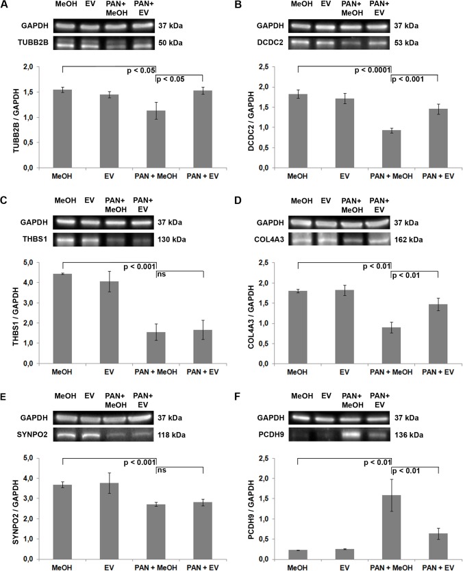
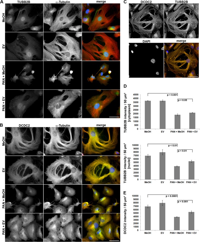
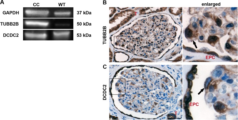
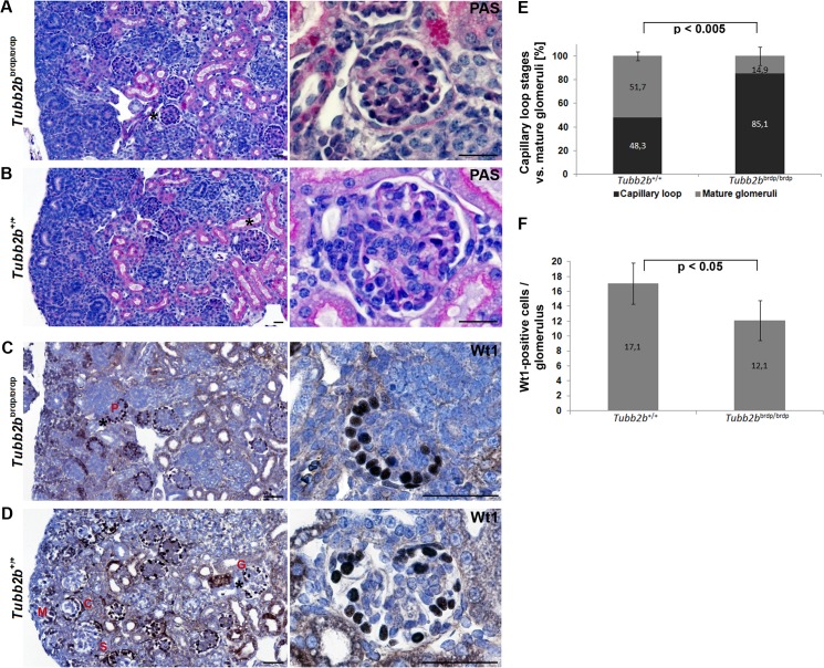
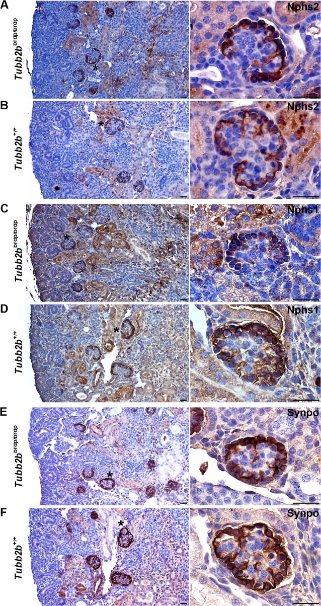
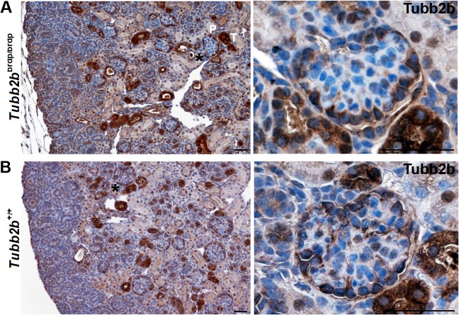
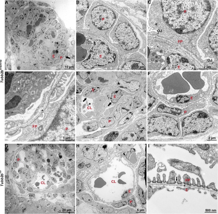
Results: Upon treatment with puromycin aminonucleoside, microarray analysis revealed gene clusters involved in cytoskeletal reorganization, cell adhesion, migration and extracellular matrix composition to be affected. Everolimus was capable of protecting podocytes from injury, both on transcriptional and protein level. Rescued genes included tubulin beta 2B class IIb (TUBB2B) and doublecortin domain containing 2 (DCDC2), both involved in microtubule structure formation in neuronal cells but not identified in podocytes so far. Validating gene expression data, Western-blot analysis in cultured podocytes demonstrated an increase of TUBB2B and DCDC2 protein after everolimus treatment, and immunohistochemistry in healthy control kidneys confirmed a podocyte-specific expression. Interestingly, Tubb2bbrdp/brdp mice revealed a delay in glomerular podocyte development as showed by podocyte-specific markers Wilm's tumour 1, Podocin, Nephrin and Synaptopodin.

Conclusions: Taken together, our study suggests that off-target, non-immune mediated effects of the mTOR-inhibitor everolimus on the podocyte cytoskeleton might involve regulation of microtubules, revealing a potential novel role of TUBB2B and DCDC2 in glomerular podocyte development.


Conflict of interest statement

Competing Interests: The authors have declared that no competing interests exist.

Figures

Fig 1
Fig 1. Western-blot analysis of human podocytes: Verification of selected microarray results.
Quantification of total TUBB2B (A), DCDC2 (B), THBS1 (C), COL4A3 (D), SYNPO2 (E) and PCDH9 (F) protein. GAPDH = loading control. For quantification, total protein was normalized with respect to GAPDH (representative example from 3 independent experiments). PAN = puromycin aminonucleoside. EV = everolimus. MeOH = methanol, solvent for EV. p = p-value. Data are means ± SD. ns = not significant.
Fig 2
Fig 2. Immunofluorescence analysis of human podocytes: Verification of selected microarray results.
(A+B) alpha-tubulin, TUBB2B and DCDC2 images are presented in gray scale for maximum contrast. The merge image depicts TUBB2B and DCDC2 in green and alpha-tubulin in red. DAPI was used to visualize nuclei (blue). PAN-treated cells are smaller compared to control cells, due to alterations of the actin cytoskeleton and cell-substrate adhesion dynamics [7]. (C) Double staining of TUBB2B (red) and DCDC2 (green) of untreated control cells (D+E) Quantification of TUBB2B- and DCDC2 staining intensity with Image J software (n = 3 experiments, ≥ 10 cells per condition). PAN = puromycin aminonucleoside. EV = everolimus. MeOH = methanol, solvent for EV. p = p-value. Data are means ± SD. Scale bar = 50 μm.
Fig 3
Fig 3. TUBB2B and DCDC2 are expressed in human kidneys.
(A) Western-blot analysis to measure the existence of TUBB2B and DCDC2 in human kidneys. GAPDH = loading control. CC = Podocyte cell culture. WT = Wild type kidney. (B+C) Immunohistochemical staining of TUBB2B and DCDC2 in healthy human kidneys. 3,3'-diaminobenzidine (DAB) was used as chromogen (brown staining) and nuclei were stained with hematoxylin (blue). The black arrows mark a nuclear as well as cytoplasmic staining of TUBB2B and a cytoplasmic staining of DCDC2 in podocytes. A negative control was performed without the primary antibody (S1 Fig). T = tubule. EPC = parietal epithelial cell. Scale bar = 50 μm.
Fig 4
Fig 4. Periodic acid-Schiff and Wilm’s tumour 1 staining of wild type and Tubb2b brdp/brdp mouse kidneys (E18.5).
(A+B) Glomeruli were stained with periodic acid-Schiff to highlight basement membranes of glomerular capillary loops and tubular epithelium. (A) Tubb2b brdp/brdp mice often show a lack in glomerular tuft and capillary lumen development. (B) The capillary loops of the wild type glomeruli are well-defined and thin. Scale bars = 25 μm. (C+D) Wilm’s tumour 1 (Wt1) expression was detectable by immunohistochemistry (dark-brown nuclear staining results from 3,3'-diaminobenzidine). Nuclei were stained with hematoxylin (blue). (C) Tubb2b brdp/brdp kidneys show a specific Wt1 staining in podocytes (P) arranged in a string of pearls-like pattern at the periphery of the glomerulus, characteristic of an early developmental stage. (D) In the developing wild type kidney, Wt1 is expressed in mesenchymal cells that are starting the mesenchymal-to-epithelial transition (condensing metanephric mesenchyme (M)), in early epithelial structures (comma- (C) and S-shaped (S) bodies) and in fully differentiated epithelial cells (glomerular podocytes (G)). Black asterisks: Enlarged section areas on the right. Scale bars = 50 μm. (E) Percentage of capillary loop stages versus mature glomeruli in 3 mutant and 4 wild type animals. For each animal, 15 to 30 capillary loop stages / mature glomeruli were counted. Data are means ± SD. (F) Average number of Wt1-positive cells per glomerulus in 3 mutant and 4 wild type animals. 15 to 30 glomeruli / animal were counted. Data are means ± SD.
Fig 5
Fig 5. Nphs2-, Nphs1- and Synpo staining of wild type and Tubb2b brdp/brdp mouse kidneys (E18.5).
Immunohistochemical staining of mice kidneys (dark-brown Nphs2, Nphs1 and Synpo stainings result from 3,3'-diaminobenzidine. Nuclei were stained with hematoxylin (blue). (A+B) Nphs2 staining. (A) Tubb2b brdp/brdp kidneys show a specific Nphs2 staining in podocytes arranged like a row of pearls at the periphery of the glomerulus. (B) Within the developing wild type kidney early capillary loop stages and maturing glomeruli are labeled. (C+D) Nphs1 staining. (E+F) Synpo staining. (C-F) Both, Nphs1 and Synpo patterns are comparable to the Nphs2 staining. Black asterisks: Enlarged section areas on the right. Scale bars = 50μm.
Fig 6
Fig 6. Tubb2b staining of wild type and Tubb2b brdp/brdp mouse kidneys (E18.5).
Immunohistochemical staining of mice kidneys (dark-brown Tubb2b staining results from 3,3'-diaminobenzidine. Nuclei were stained with hematoxylin (blue). (A) Tubb2b brdp/brdp kidneys show a specific cytoplasmic Tubb2b expression in tubuli (T) and in podocytes (P), confirming the developmental defects seen with the Wt1, Nphs2, Nphs1 and Synpo stainings. (B) Tubb2b expression in wild type kidneys is restricted to the mature podocytes. Interestingly in the developing wild type kidney Tubb2b is not expressed in the early developmental stages of maturing podocytes. Note, that in murine podocytes nuclear Tubb2b seems much less expressed compared to human kidneys. Black asterisks: Enlarged section areas on the right. Scale bars = 50 μm.
Fig 7
Fig 7. Transmission electron micrographs of wild type and Tubb2b brdp/brdp mouse kidneys (E18.5).
(A-F) With TEM, a delay in glomerular endothelial (E) and podocyte (P) development could be observed in Tubb2b brdp/brdp mice. (A+B) Glomerulus with immature, cuboidal podocytes (black arrow). (C+D) Incompletely differentiated podocyte foot processes (FPs); some are extremely wide and linked by occludens junctions (OJs). (D) The glomerular basement membrane (asterisk) appears to be normal. (A+E) Glomeruli with no or small visible capillary lumen (CL) and multiple endothelial cells within the capillary loops (black arrows). (F) Swollen and vacuolated glomerular endothelial cells with decreased fenestrations. (G-I) Glomeruli of wild type Tubb2b mice had open glomerular capillaries with fenestrated endothelium, differentiated podocyte foot processes linked by SDs (black arrow) and a normal glomerular basement membrane.

References

    1. Mundel P, Kriz W. Structure and function of podocytes: an update. Anat Embryol (Berl). 1995; 192(5): 385–397. - PubMed
    1. Pavenstadt H, Kriz W, Kretzler M. Cell biology of the glomerular podocyte. Physiol Rev. 2003; 83(1): 253–307. - PubMed
    1. Yuan H, Takeuchi E, Salant DJ. Podocyte slit-diaphragm protein nephrin is linked to the actin cytoskeleton. Am J Physiol Renal Physiol. 2002; 282(4): F585–F591. - PubMed
    1. Büscher AK, Weber S. Educational paper: the podocytopathies. Eur J Pediatr. 2012; 171(8): 1151–1160. 10.1007/s00431-011-1668-2 - DOI - PubMed
    1. Takai Y, Sasaki T, Matozaki T. Small GTP-binding proteins. Physiol Rev. 2001; 81(1): 153–207. - PubMed

Publication types

Associated data