. 2015 Sep 2;10(9):e0136951.
doi: 10.1371/journal.pone.0136951. eCollection 2015.

Altered FGF Signaling Pathways Impair Cell Proliferation and Elevation of Palate Shelves

Affiliations

Altered FGF Signaling Pathways Impair Cell Proliferation and Elevation of Palate Shelves

Weijie Wu et al. PLoS One. .

Abstract

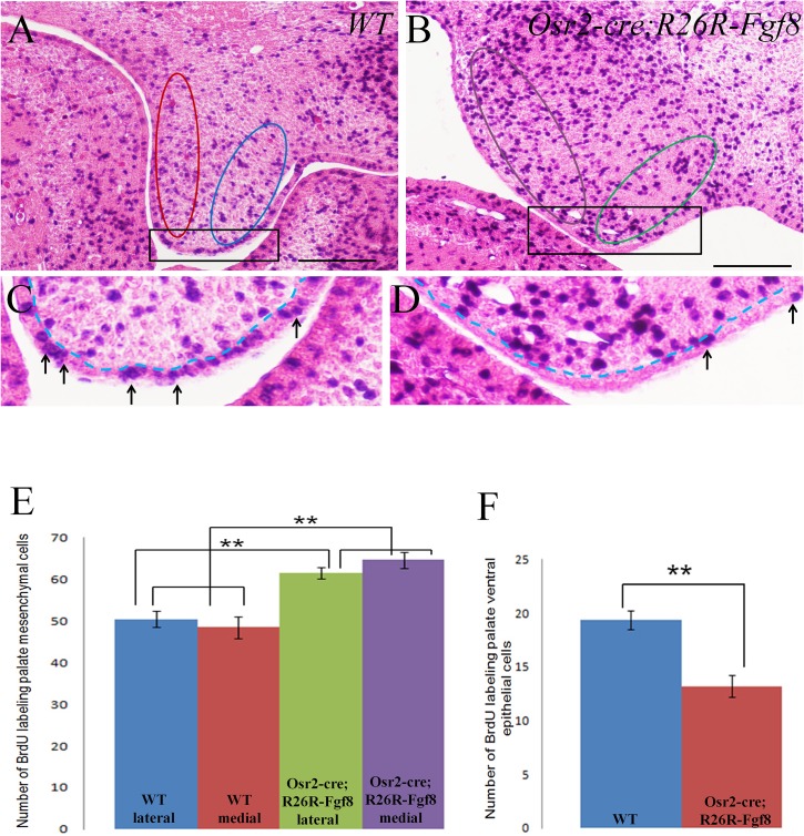
In palatogenesis, palatal shelves are patterned along the mediolateral axis as well as the anteroposterior axis before the onset of palatal fusion. Fgf10 specifically expressed in lateral mesenchyme of palate maintains Shh transcription in lateral epithelium, while Fgf7 activated in medial mesenchyme by Dlx5, suppressed the expansion of Shh expression to medial epithelium. How FGF signaling pathways regulate the cell behaviors of developing palate remains elusive. In our study, we found that when Fgf8 is ectopically expressed in the embryonic palatal mesenchyme, the elevation of palatal shelves is impaired and the posterior palatal shelves are enlarged, especially in the medial side. The palatal deformity results from the drastic increase of cell proliferation in posterior mesenchyme and decrease of cell proliferation in epithelium. The expression of mesenchymal Fgf10 and epithelial Shh in the lateral palate, as well as the Dlx5 and Fgf7 transcription in the medial mesenchyme are all interrupted, indicating that the epithelial-mesenchymal interactions during palatogenesis are disrupted by the ectopic activation of mesenchymal Fgf8. Besides the altered Fgf7, Fgf10, Dlx5 and Shh expression pattern, the reduced Osr2 expression domain in the lateral mesenchyme also suggests an impaired mediolateral patterning of posterior palate. Moreover, the ectopic Fgf8 expression up-regulates pJak1 throughout the palatal mesenchyme and pErk in the medial mesenchyme, but down-regulates pJak2 in the epithelium, suggesting that during normal palatogenesis, the medial mesenchymal cell proliferation is stimulated by FGF/Erk pathway, while the epithelial cell proliferation is maintained through FGF/Jak2 pathway.


Conflict of interest statement

Competing Interests: The authors have declared that no competing interests exist.

Figures

Fig 1
Fig 1. Cre pattern in the developing palatal shelves detected by lacZ expression in Osr2-Cre KI ; Rosa26R-LacZ embryos. (A-F)
Cre-mediated LacZ expression in the developing Osr2-Cre KI ; Rosa26R-LacZ palatal shelves. At E11.5, both the nascent anterior (A) and posterior palatal shelves (B) were devoid of LacZ staining. A slight LacZ staining was only detected in the connecting area between mandible and maxillary process (Arrows in A). At E12.5, Cre-mediated LacZ expression was activated throughout the anterior (C) and posterior palatal mesenchyme (D), but specifically absent from the palatal epithelium (arrows in C&D). At E13.5, Cre activity evenly distributed anterior palate mesenchyme (E). However, Cre activity was only found in the medial and lateral mesenchyme of the posterior palate (arrowheads in F), but absent from the central part (* in F). Judged by the LacZ intensity, there was no difference in Cre activity between the medial and lateral palatal mesenchyme (C-F). (Scale bar: 200um)
Fig 2
Fig 2. The complete cleft palate in Osr2-Cre KI ; Rosa26R-Fgf8 mouse embryos.
(A-L) Cross sections by HE staining of the developing Osr2-Cre KI ; Rosa26R-Fgf8 palatal shelves. At E13.5, the anterior palatal shelves showed no difference between the WT (A) and Osr2-Cre KI ; Rosa26R-Fgf8 (B) embryos. By contrast, the mutant posterior palatal shelves (D) were significantly enlarged in size, compared with the WT posterior palatal shelves (C), especially in the medial side (bidirectional arrow in D). When the WT palatal shelves elevated horizontally at the anterior (E) and even initiated fusion by forming middle epithelial seam in the posterior (arrow in G) at E14.5, the Osr2-Cre KI ; Rosa26R-Fgf8 palatal shelves still kept their vertical status in both anterior (F) and the posterior (H) with the obvious enlarged medial side (bidirectional arrow in H). At E18.5, the Osr2-Cre KI ; Rosa26R-Fgf8 embryos exhibited the complete cleft palate: the anterior palatal shelves only elevated slightly (J) and the enlarged posterior shelves showed no sign of elevation (L, bidirectional arrow delineated the enlarged medial mesenchyme). By contrast, the E18.5 WT palate shelves had fused with each other and separated oral from nasal cavity in the anterior (I) and posterior (K) (Scale bar: 200um)
Fig 3
Fig 3. The examination on cell proliferation ratio of the developing palatal shelves of Osr2-Cre KI ; Rosa26R-Fgf8 mouse embryos.
(A-D) Cell proliferation test by BrdU labelling for the E13.5 Osr2-Cre KI ; Ros26R-Fgf8 palatal shelves. The BrdU labelled cells in the E13.5 WT medial (red circle in A) and lateral palatal mesenchyme (blue circle in A) were less than those in the Osr2-Cre KI ; Ros26R-Fgf8 medial (violet circle in B) and lateral mesenchyme (green circle in B). Opposing to the mesenchymal cell proliferation ratio, the number of BrdU positive cells in the E13.5 WT palatal epithelium (arrows in C) was more than that in the mutant palatal epithelium (arrows in D). (E, F) Statistics of the numbers of BrdU labelled cells in the E13.5 palatal shelves. The number of proliferating medial mesenchymal cells significantly raised from 48.455 (SD = 8.722, SE = 2.629) in WT to 64.667 (SD = 5.937, SE = 1.979) in the Osr2-Cre KI ; Rosa26R-Fgf8 mouse (P<0.01). Similarly, the proliferating cells in lateral mesenchyme raised greatly from 50.455 (SD = 6.758, SE = 1.983) in WT to 61.556 (SD = 4.362, SE = 1.454) in the Osr2-Cre KI ; Rosa26R-Fgf8 mouse (P<0.01) (E). The difference in cell proliferation ratio between the medial and lateral palatal mesenchyme had no significance in both WT and Osr2-Cre KI ; Rosa26R-Fgf8 mouse (E). Reversely, the amount of proliferating cells in palatal epithelium dropped drastically from 19.364 (SD = 2.942, SE = 0.887) in WT to 13.322 (SD = 3.114, SE = 1.038) in the Osr2-Cre KI ; Ros26R-Fgf8 mouse (P<0.01) (F). (C and D were the enlarged areas of the square boxes in A and B, respectively; Dashed lines in C and D delineated the boundary between mesenchyme and epithelium; SD, Standard Derivation; SE, Standard Error; Scale bar: 200um)
Fig 4
Fig 4. FGF8 does not directly inhibit Shh expression and palatal epithelial cell proliferation.
(A-D) Whole mount in situ hybridization for Shh expression in Osr2-Cre KI ;Rosa26R-Fgf8 palate. At E13.5, a robust Shh transcription was detected in the most anterior rugae of the WT palate (arrows in A); only a trace of Shh transcription was found in the most anterior ruga in mutant littermate (arrow in B). At E14.5, there were 8 Shh-expressing rugae in WT palate (arrows in C), while in Osr2-Cre KI ;Rosa26R-Fgf8 palate, only two short Shh-expressing rugae were emerging (arrows in D). (E, F) Whole mount in situ hybridization with Shh probe in Shh-Cre;Rosa26R-Fgf8 palate. Although a complete cleft palate was detected in Shh-Cre;Rosa26R-Fgf8 mouse at E14.5 (F), both the WT (E) and their mutant littermates (F) show 8 Shh-expressing rugae. (G) X-gal staining demonstrates that at E13.5, Cre activity was distributed all over the palatal epithelium of the Shh-Cre;Rosa26R-LacZ mouse. (H-K) BrdU labelled test and statistical analysis for E13.5 palatal cell proliferation. The E13.5 cell proliferation of WT (H) and Shh-Cre;Rosa26R-Fgf8 (I) palatal shelves showed significant difference in mesenchyme (J) and no difference in epithelium (K). The BrdU labelled cells of Shh-Cre;Rosa26R-Fgf8 lateral mesenchyme (42.625, SD = 6.805, SE = 2.404; violet column in J) were significantly less than that of WT control (53.25, SD = 3.615, SE = 1.278; red column in J) (P<0.01); similarly, the BrdU positive cells in medial Shh-Cre;Rosa26R-Fgf8 mesenchyme (49, SD = 4.44, SE = 1.569; green column in J) were dramatically less than WT littermate (55.625, SD = 3.335, SE = 1.179; blue column in J) (P<0.01). Although the cell proliferation of both the medial and lateral Shh-Cre;Rosa26R-Fgf8 mesenchyme decreased, the drop in the lateral side was more significant than the medial side (P<0.05). The BrdU labelled epithelial cells in Shh-Cre;Rosa26R-Fgf8 palate (22.5, SD = 2.976, SE = 1.052; red column in K) were slightly more than WT control (21, SD = 3.023,SE = 1.069; blue column in K), but the increase has no significance (P>0.05) (Red and blue circles in H and violet and green circles in I delineate medial and lateral mesenchyme, respectively; Dashed lines in B, D and F delineate the edge of palatal shelves; Dashed lines in H and I stand for the layer of epithelium; SD, Standard Deviation;SE, Standard Error; Scale bar: 200um)
Fig 5
Fig 5. The expression pattern ofmesenchyme markers inthe Osr2-Cre KI ;Rosa26R-Fgf8 palate.
(A-J) In situ hybridization for mesenchymal markers of E13.5 palatal shelves. Fgf10 was activated in the lateral mesenchyme of WT palate (arrow in A), but silenced in Osr2-Cre KI ;Rosa26R-Fgf8 palate (B). Similarly, Fgf7 activated in WT medial palatal mesenchyme (arrow in C) disappeared in mutant palate (D). The expression of Dlx5 acting as the activator of Fgf7 was detected in medial side of WT palatal mesenchyme (arrow in E), but not in the palate of mutant littermate (F). Compared with the Osr2 expression in WT lateral palate (arrow in G), the domain of Osr2 in the Osr2-Cre KI ;Rosa26R-Fgf8 palate reduced from the distal mesenchyme (arrow in H). The expression of Wnt5a detected in lateral mesenchyme of the Osr2-Cre KI ;Rosa26R-Fgf8 palate (arrow in J) was comparable to that in the WT (arrow in I).
Fig 6
Fig 6. The pattern ofphosphorylated FGF signaling mediators in the Osr2-Cre KI ;Rosa26R-Fgf8 palate.
(A-H) Immunohistochemistry for phosphorylated FGF signaling mediators in E13.5 palatal shelves. pErk was specifically activated in the medial mesenchyme of Osr2-Cre KI ; Rosa26R-Fgf8 palate (arrow in B), but absent from WT palate mesenchyme (A). WT palatal mesenchyme was devoid of pJak1 (C), while the mutant palatal mesenchyme showed a strong level of pJak1 (D). Normally, the pJak2 was limited to the WT lateral mesenchyme (arrow in E) and absent from palatal epithelium (E); by contrast, the pJak2 was diminished in Osr2-Cre KI ; Rosa26R-Fgf8 palatal mesenchyme and activated in the epithelium (F). The level of pAkt in the mesenchyme and epithelium of WT palate (G) is comparable to that in mutant palate (H). Similarly, compared with the WT control (I), the level of pPLCγ1 showed no difference in Osr2-Cre KI ; Rosa26R-Fgf8 palatal mesenchyme and epithelium (J). (A’-F’) The enlarged views of the boxed areas in A-F. Black arrow in B’ meant the enhanced pErk area; black arrow in E’ pointed to the active pJak2 region, and red arrows in E’ and F’ to palatal epithelium; the dashed line in E and E’ delineated the boundary between medial and lateral mesenchyme. (Scale bar: 200um)

Similar articles

Cited by

References

    1. Bush JO, Jiang RL. (2012) Palatogenesis: morphogenetic and molecular mechanisms of secondary palate development. Development. 139: 231–243. 10.1242/dev.067082 - DOI - PMC - PubMed
    1. Dudas M, Li WY, Kim J, Yang A, Kaartinen V. (2007) Palatal fusion—where do the midline cells go? A review on cleft palate, a major human birth defect. Acta Histochem. 109:1–14. - PubMed
    1. Gritli-Linde A. (2007) Molecular control of secondary palate development. Dev Biol. 301:309–326. - PubMed
    1. He FL, Xiong W, Yu X, Espinoza-Lewis R, Liu C, Gu S, et al. (2008) Wnt5a regulates directional cell migration and cell proliferation via Ror2-mediated non-canonical pathway in mammalian palate development. Development. 135: 3871–3879. 10.1242/dev.025767 - DOI - PMC - PubMed
    1. Welsh IC, O'Brien TP. (2009) Signaling integration in the rugae growth zone directs sequential SHH signaling center formation during the rostral outgrowth of the palate. Dev Biol. 336: 53–67 10.1016/j.ydbio.2009.09.028 - DOI - PMC - PubMed

Publication types