Review
. 2016 Aug 15;594(16):4565-79.
doi: 10.1113/JP270590. Epub 2015 Oct 14.

Membrane lipid rafts and neurobiology: age-related changes in membrane lipids and loss of neuronal function

Affiliations
Review

Membrane lipid rafts and neurobiology: age-related changes in membrane lipids and loss of neuronal function

Junji Egawa et al. J Physiol. .

Abstract

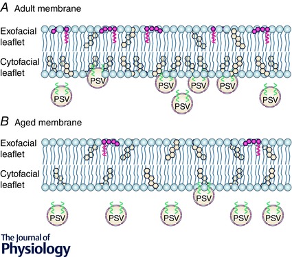
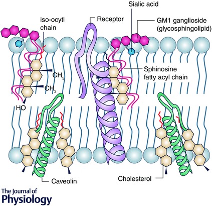
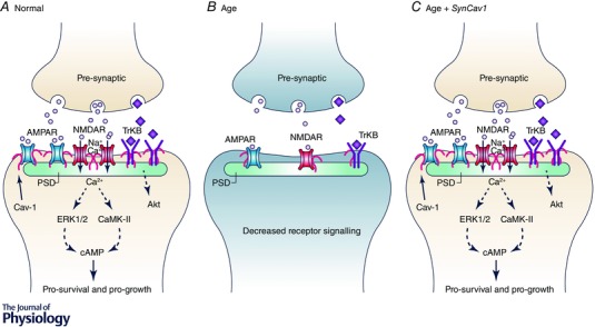
A better understanding of the cellular physiological role that plasma membrane lipids, fatty acids and sterols play in various cellular systems may yield more insight into how cellular and whole organ function is altered during the ageing process. Membrane lipid rafts (MLRs) within the plasma membrane of most cells serve as key organizers of intracellular signalling and tethering points of cytoskeletal components. MLRs are plasmalemmal microdomains enriched in sphingolipids, cholesterol and scaffolding proteins; they serve as a platform for signal transduction, cytoskeletal organization and vesicular trafficking. Within MLRs are the scaffolding and cholesterol binding proteins named caveolin (Cav). Cavs not only organize a multitude of receptors including neurotransmitter receptors (NMDA and AMPA receptors), signalling proteins that regulate the production of cAMP (G protein-coupled receptors, adenylyl cyclases, phosphodiesterases (PDEs)), and receptor tyrosine kinases involved in growth (Trk), but also interact with components that modulate actin and tubulin cytoskeletal dynamics (e.g. RhoGTPases and actin binding proteins). MLRs are essential for the regulation of the physiology of organs such as the brain, and age-related loss of cholesterol from the plasma membrane leads to loss of MLRs, decreased presynaptic vesicle fusion, and changes in neurotransmitter release, all of which contribute to different forms of neurodegeneration. Thus, MLRs provide an active membrane domain that tethers and reorganizes the cytoskeletal machinery necessary for membrane and cellular repair, and genetic interventions that restore MLRs to normal cellular levels may be exploited as potential therapeutic means to reverse the ageing and neurodegenerative processes.


Figures

Figure 1
Figure 1. Aged‐related changes in cholesterol and gangliosides in the neuronal plasma membrane
A, in young adult human and rodent neurons, approximately 85% of plasmalemmal cholesterol is found within the cytofacial leaflet while the majority of glycosphingolipids (e.g. GM1 gangliosides) are found in the exofacial leaflet. Both gangliosides and cholesterol enhance the negative curvature property of the plasma membrane, thus enhancing the fusogenicity with presynaptic vesicles (PSV). B, however, with age there is either a reduction in membrane gangliosides and cholesterol or a redistribution of cholesterol from the cyto‐ to the exofacial leaflet. These age‐related changes in cholesterol membrane content and distribution drastically impede PSV docking and fusion with the cytofacial leaflet, neurotransmitter release and postsynaptic signalling (e.g. NMDAR and AMPAR), synaptic plasticity (i.e. LTP) and strength, and behaviour.
Figure 2
Figure 2
The physical interaction between cholesterol, sphingolipids and transmembrane proteins Cholesterol physically interacts with sphingolipids and the transmembrane component of proteins. Cholesterol possesses an α smooth surface, which permits it to interact with sphingolipids, while the β rough surface (due to methyl groups on carbon 10 and 13 and the iso‐octyl chain link to carbon 17) facilitates insertion into the helices of transmembrane proteins. Due to its dissymmetrical physical properties, one molecule of cholesterol can interact with two distinct membrane molecules, such as sphingolipids and transmembrane proteins, within raft microdomains. This serves to create a more liquid ordered membrane parameter (i.e. decreased fluidity) characteristic of MLR.
Figure 3
Figure 3. Age‐related loss in synaptic signalling may be restored by increasing MLR formation
A, in the healthy brain, synaptic transmission is dependent upon the appropriate neuronal receptors (e.g. AMPAR, NMDAR, and TrkB), their localization to the postsynaptic density, and release of neurotransmitter from the presynaptic cleft. MLRs, which are critical membrane components of both the pre‐ and postsynaptic membrane, scaffold these receptors in part via the cholesterol binding protein Cav‐1. B, however, with age there is a drastic decrease in neuronal MLRs, Cav‐1, and MLR‐associated neuronal receptors, which results in decreased synaptic transmission (reduced cAMP formation), progressive neurodegeneration, and increased behavioural dysfunction. C, most highly targeted therapies are often ineffective in eliciting the desired response, likely to be due to decreased expression of key receptors (e.g. AMPARs, NMDARs, TrkB, GPCRs) and inadequate production of second messenger (cAMP). Genetic interventions that enhance and/or restore MLR formation (i.e. neuron‐targeted Cav‐1 or SynCav1) can potentially re‐establish an active signalling platform that regulates cytoskeletal dynamics, enhances structural and functional neuroplasticity, and improves cognition in the ageing and neurodegenerative brain.

References

    1. Allen JA, Halverson‐Tamboli RA & Rasenick MM (2007). Lipid raft microdomains and neurotransmitter signalling. Nat Rev Neurosci 8, 128–140. - PubMed
    1. Atkins CM, Cepero ML, Kang Y, Liebl DJ & Dietrich WD (2013). Effects of early rolipram treatment on histopathological outcome after controlled cortical impact injury in mice. Neurosci Lett 532, 1–6. - PMC - PubMed
    1. Atkins CM, Falo MC, Alonso OF, Bramlett HM & Dietrich WD (2009). Deficits in ERK and CREB activation in the hippocampus after traumatic brain injury. Neurosci Lett 459, 52–56. - PMC - PubMed
    1. Atkins CM, Oliva AA Jr, Alonso OF, Pearse DD, Bramlett HM & Dietrich WD (2007). Modulation of the cAMP signaling pathway after traumatic brain injury. Exp Neurol 208, 145–158. - PMC - PubMed
    1. Aziz R & Steffens DC (2013). What are the causes of late‐life depression? Psychiatr Clin North Am 36, 497–516. - PMC - PubMed

Publication types