

Tarsal tunnel syndrome masked by painful diabetic polyneuropathy
- PMID: 26333036
- PMCID: PMC4601976
- DOI: 10.1016/j.ijscr.2015.08.033
Tarsal tunnel syndrome masked by painful diabetic polyneuropathy
Abstract
Introduction: Various causes influence the etiology of tarsal tunnel syndrome including systemic diseases with progressive neuropathy, such as diabetes.
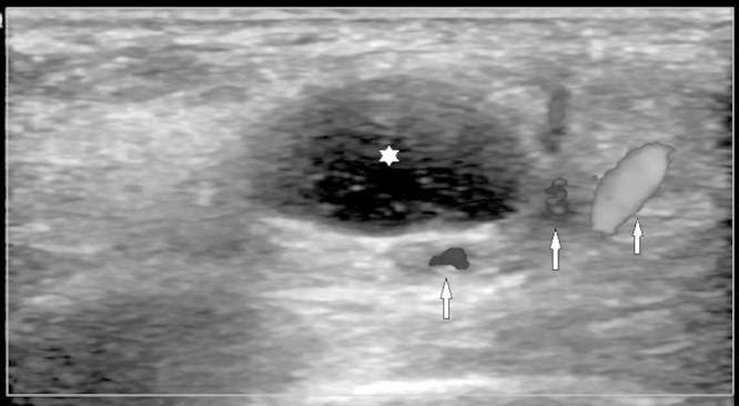
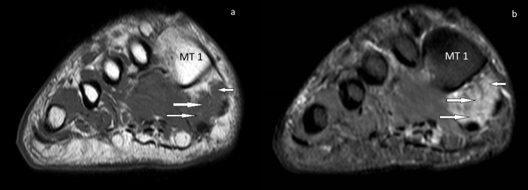
Presentation of case: We describe a 52-year-old male patient with complaints of numbness, burning sensation and pain in both feet. The laboratory results showed that the patient had uncontrolled diabetes, and the EMG showed distal symmetrical sensory-motor neuropathy and nerve entrapment at the right. Ultrasonography and MRI showed the cyst in relation to medial plantar nerve, and edema- moderate atrophy were observed at the distal muscles of the foot.
Discussion: Foot neuropathy in diabetic patients is a complex process. So, in planning the initial treatment, medical or surgical therapy is selected based on the location and type of the pathology. Foot deformities can be corrected with resting, anti-inflammatory treatment, appropriate shoes, orthesis and socks, and if required, ankle stabilization can be attempted. If the patient is still unresponsive, surgical treatment may be applied.
Conclusion: It is essential to investigate more localized reasons like tarsal tunnel syndrome that may mimic diabetic neuropathy, should be treated primarily.
Keywords: Diabetic polyneuropathy; Magnetic resonance; Pain; Superficial ultrasound; Tarsal tunnel syndrome.
Copyright © 2015 The Authors. Published by Elsevier Ltd.. All rights reserved.
Figures


References
-
- Delfaut E.M., Demondion X., Bieganski A., Thiron M.C., Mestdagh H., Cotten A. Imaging of foot and ankle nerve entrapment syndromes: from well-demonstrated to unfamiliar sites. Radiographics. 2003;23:613–623. - PubMed
-
- Aszmann O.C., Ebmer J.M., Dellon A.L. Cutaneous innervation of the medial ankle: an anatomic study of the saphenous, sural, and tibial nerves and their clinical significance. Foot Ankle Int. 1998;19:753–756. - PubMed
-
- Sammarco G.J., Conti S.F. Tarsal tunnel syndrome caused by an anomalous muscle. J. Bone Joint Surg. Am. 1994;76:1308–1314. - PubMed
-
- Trepman E., Kadel N.J., Chisholm K., Razzano L. Effect of foot and ankle position on tarsal tunnel compartment pressure. Foot Ankle Int. 1999;20:721–726. - PubMed
-
- Donovan A., Rosenberg Z.S., Cavalcanti C.F. MR imaging of entrapment neuropathies of the lower extremity. Part 2. The knee, leg, ankle, and foot. Radiographics. 2010;30(4):1001–1019. - PubMed
LinkOut - more resources
Full Text Sources
Other Literature Sources
Research Materials
