. 2015 Sep 3:6:8143.
doi: 10.1038/ncomms9143.

A highly stable prefusion RSV F vaccine derived from structural analysis of the fusion mechanism

Affiliations

A highly stable prefusion RSV F vaccine derived from structural analysis of the fusion mechanism

Anders Krarup et al. Nat Commun. .

Abstract

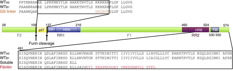
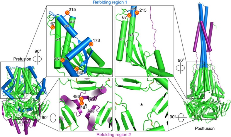
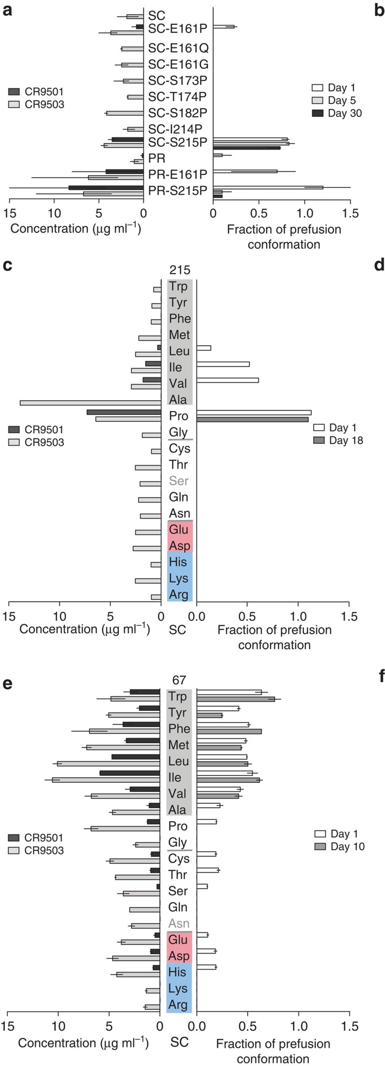
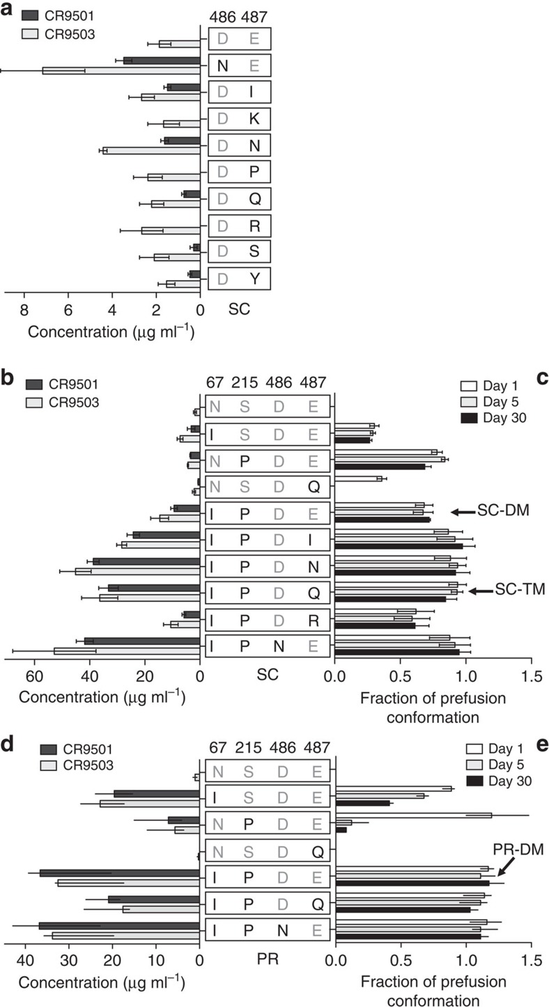
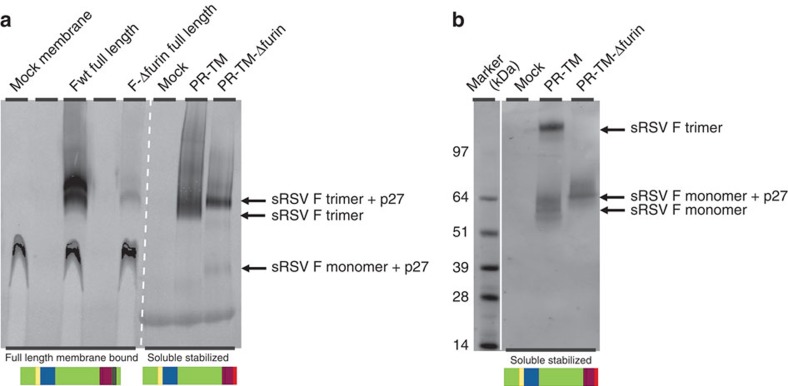
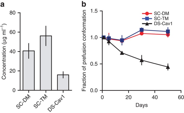
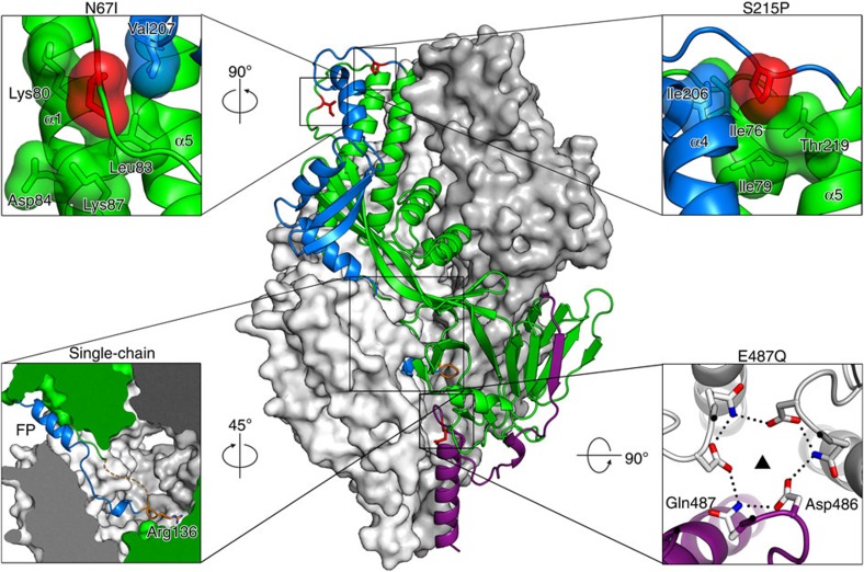
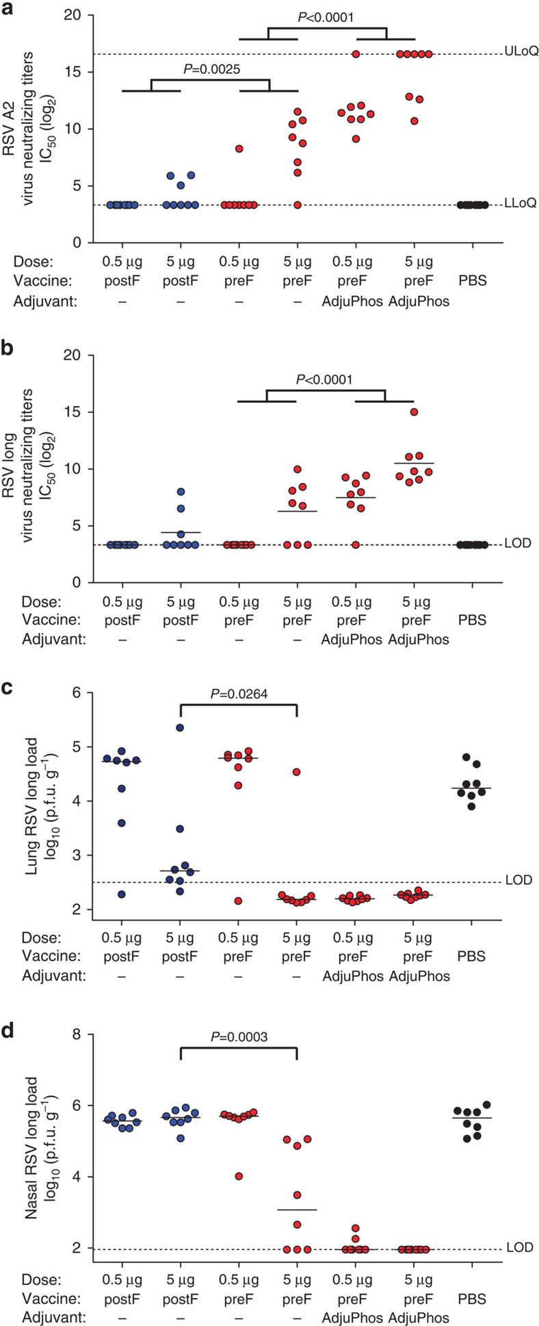
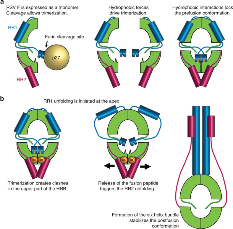
Respiratory syncytial virus (RSV) causes acute lower respiratory tract infections and is the leading cause of infant hospitalizations. Recently, a promising vaccine antigen based on the RSV fusion protein (RSV F) stabilized in the native prefusion conformation has been described. Here we report alternative strategies to arrest RSV F in the prefusion conformation based on the prevention of hinge movements in the first refolding region and the elimination of proteolytic exposure of the fusion peptide. A limited number of unique mutations are identified that stabilize the prefusion conformation of RSV F and dramatically increase expression levels. This highly stable prefusion RSV F elicits neutralizing antibodies in cotton rats and induces complete protection against viral challenge. Moreover, the structural and biochemical analysis of the prefusion variants suggests a function for p27, the excised segment that precedes the fusion peptide in the polypeptide chain.


Figures

Figure 1
Figure 1. Schematic representation of RSV F0 with refolding region 1 (RR1 in blue), refolding region 2 (RR2 in purple), p27 (yellow) and remainder (green).
F0 protein is cleaved at two positions (arrows) to generate F1 and F2. Upper sequence alignment shows design of single-chain (SC) with short linker region (orange) compared with fragment of Fwt of strain A2 (subgroup A) and B1 (subgroup B). Lower sequence alignment shows design of soluble truncated variant and variant with fibritin domain (red) compared with fragment of Fwt of A2 and B1.
Figure 2
Figure 2. Strategy for prefusion F protein stabilization.
RSV F trimer and magnified views of prefusion F (left) and postfusion F protein (right) indicating refolding region 1 (RR1, blue) and refolding region 2 (RR2, purple) and location of amino acid substitutions indicated by crossed circles. The substitutions in the RR1 are designed to prevent the formation of the long helix. The substitutions in the RR2 are designed to reduce the negative charge repulsion between the β23 strands.
Figure 3
Figure 3. Amino acid substitutions at positions 67 and 215 increase expression level and prefusion stability of the F protein.
(a,b) Protein expression levels and prefusion stability of RSV F SCA2 variants and processed variants (PRA2) with substitutions in RR1 (n=2–4). (c,d) Protein expression levels and prefusion stability of RSV F SCA2 variants with all 20 amino acid substitutions at position 215 (n=1) and (e,f) at position 67 (n=2). Protein expression levels in cell culture supernatants were tested 72 h post transfection and fraction of RSV F protein binding to prefusion specific CR9501 antibody on the day of harvest and after storage at 4 °C for indicated period of time. (a,b,e and f)—bars represent average of 2–4 measurements, lines represent range of values; (c,d)—bars represent single measurement. Amino acids are grouped according to physicochemical characteristics (grey: hydrophobic, red: negative charge, blue: positive charge). Variants in this and next figures are based on strain A2.
Figure 4
Figure 4. Amino acid substitutions have additive effect on RSV F protein expression and stability.
(a) Protein expression levels of RSV F SCA2 variants with substitutions at position 486 and 487. (b,c) Protein expression levels and prefusion stability of RSV F SCA2 variants with multiple substitutions. (d,e) Protein expression levels and prefusion stability of processed RSV F PRA2 variants with multiple amino acid substitutions. Protein expression levels in cell culture supernatants were tested 72 h post transfection and fraction of RSV F protein binding to prefusion-specific CR9501 antibody on the day of harvest and after storage at 4 °C for indicated period of time. (ac) n=3–4, Mean±s.e.; (d,e) bars represent average of 2–4 measurements, lines represent range of values.
Figure 5
Figure 5. Influence of p27 on trimerization.
(a, left panel) Full-length RSV F A2 and a variant mutated in one of the furin cleavage sites (F-ΔFurin: R135Q, R136Q) were expressed in HEK293T cells and the purified membrane fractions were analysed by NativePAGE followed by Western blot to evaluate expression and quaternary structure. (a, right panel) Soluble, stabilized processed F variant with fibritin motif (PR-TM: N67I, S215P, D486N) and a furin site cleavage mutant (PR-TM-ΔFurin) were analysed on NativePAGE followed by Western blot and (b) SDS–PAGE followed by Western blot. For SDS-PAGE, samples were treated with standard 0.1% SDS buffer without reducing agents and without boiling. (bottom) Schematic representations of RSV F with identical colour coding as Fig. 1 with additional transmembrane region (dark green) and fibritin domain (red).
Figure 6
Figure 6. Comparison to DS-Cav1.
(a) Protein expression levels for double mutant single chain (SC-DM) and triple mutant single chain (SC-TM) were compared with DS-Cav1 (ref. 10) in cell culture supernatants 72 h post transfection and (b) the fraction of RSV F protein binding to prefusion-specific CR9501 antibody after storage at 4 °C for indicated days. Bars or symbols represent average from two measurements, lines represent range of values. On (b) prefusion F fraction at day of harvest (day 0) is normalized to 1.
Figure 7
Figure 7. Crystal structure of SC-TM.
The trimer is displayed with two protomers as white and grey molecular surfaces and the third protomer as a green ribbon. RR1 with its N-terminal FP is dark blue, and RR2 is dark purple. The stabilizing mutations (Ile67, Pro215 and Gln487) are coloured red (except in the lower right panel), and the loop connecting F1 and F2 is orange. Each of the smaller panels depicts a magnified view of one of the four stabilizing strategies, and their orientation with respect to the main panel is indicated. The lower left panel shows a slice through the trimer, and the dashed orange line represents the disordered portion of the loop. In the lower right panel, hydrogen bonds are depicted as black dotted lines.
Figure 8
Figure 8. Prefusion RSV F protein is immunogenic and protective in Cotton rats.
Female Cotton rats (n=8 per group) were immunized IM with 0.5 or 5 μg stabilized prefusion RSV F protein (SC-DM) or RSV F protein in postfusion conformation in a prime—boost regimen at week 0 and week 4. SC-DM groups at the same protein doses was further combined with AdjuPhos. Serum was collected on day 49 and plaque reduction assay was performed using (a) RSV A2 and (b) Long as described in Materials and Methods. RSV virus titer was determined in (c) the lungs or (d) nose tissue homogenates of cotton rats at day 5 after challenge (day 54 after prime immunization) as determined by plaque forming units per gram of tissue. Horizontal lines indicate medians. Limit of detection is shown as dotted line. Groups were statistically compared using a non-parametric Cochran-Mantel-Haenszel test. Comparisons across doses are indicated by horizontal lines above the 0.5 and 5 μg groups. Significance was accepted at the P<0.05 level and P-values below 0.05 are indicated in the figure.
Figure 9
Figure 9. Schematic representation describing how cleavage can activate trimerization and how trimerization causes an inherent instability in the RR2.
(a) Trimerization of monomeric RSV F is obstructed by the uncleaved bulky glycosylated p27 region. Cleavage of F0 allows trimerization and hydrophobic interaction of the fusion peptide with adjacent protomer. (b) The trimerization creates an unstable area in the RR2 through the alignment of a repulsive ring of negative charges contributed by residues Asp486 and Glu487 that are located on β23 that contains an unstable β-bulge (at the edge of β-sheet 3, Fig. 2). The repulsive electrostatic and static forces are countered by the hydrophobic forces linking the FP to the bottom of the cavity. After RR1 is triggered to refold by an unknown mechanism at the apex, it pulls away the stabilizing hydrophobic FP from the cavity which will destabilize the repulsive ring. This results in release of RR2 and ultimately binding of HRB to the HRA coiled-coil to form a 6-helix bundle that is characteristic of the postfusion conformation.

References

    1. Nair H. et al.. Global burden of acute lower respiratory infections due to respiratory syncytial virus in young children: a systematic review and meta-analysis. Lancet 375, 1545–1555 (2010). - PMC - PubMed
    1. Hall C. B. et al.. The burden of respiratory syncytial virus infection in young children. N. Engl. J. Med. 360, 588–598 (2009). - PMC - PubMed
    1. Glezen W. P., Taber L. H., Frank A. L. & Kasel J. A. Risk of primary infection and reinfection with respiratory syncytial virus. Am. J. Dis. Child. 140, 543–546 (1986). - PubMed
    1. Zhou H. et al.. Hospitalizations associated with influenza and respiratory syncytial virus in the United States, 1993-2008. Clin. Infect. Dis. 54, 1427–1436 (2012). - PMC - PubMed
    1. Magro M. et al.. Neutralizing antibodies against the preactive form of respiratory syncytial virus fusion protein offer unique possibilities for clinical intervention. Proc. Natl Acad. Sci. USA 109, 3089–3094 (2012). - PMC - PubMed

Publication types

MeSH terms

Substances