. 2015 Sep 3:5:13659.
doi: 10.1038/srep13659.

Architecture of the Synaptophysin/Synaptobrevin Complex: Structural Evidence for an Entropic Clustering Function at the Synapse

Affiliations

Architecture of the Synaptophysin/Synaptobrevin Complex: Structural Evidence for an Entropic Clustering Function at the Synapse

Daniel J Adams et al. Sci Rep. .

Abstract

We have purified the mammalian synaptophysin/synaptobrevin (SYP/VAMP2) complex to homogeneity in the presence of cholesterol and determined the 3D EM structure by single particle reconstruction. The structure reveals that SYP and VAMP2 assemble into a hexameric ring wherein 6 SYP molecules bind 6 VAMP2 dimers. Using the EM map as a constraint, a three dimensional atomic model was built and refined using known atomic structures and homology modeling. The overall architecture of the model suggests a simple mechanism to ensure cooperativity of synaptic vesicle fusion by organizing multiple VAMP2 molecules such that they are directionally oriented towards the target membrane. This is the first three dimensional architectural data for the SYP/VAMP2 complex and provides a structural foundation for understanding the role of this complex in synaptic transmission.


Figures

Figure 1
Figure 1. SYP family protein sequence conservation.
ClustalW alignment of human SYP and paralogs colored by heat map conservation at each position assigned by the PRALINE server using the BLOSUM62 similarity matrix. The predicted TM helices based on hydropathy are shown above the sequence. G217 of SYP is located in TM4 and marked (*).
Figure 2
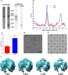
Figure 2. SYP/VAMP purification, stoichiometry and hexameric structure.
(A) Native SYP and SYP/VAMP2 complex was purified from calf brain and analyzed by SDS-PAGE, silver stain and western blot. (B) Densitometry traces of protein bands from purification without cholesterol (red) or with cholesterol (blue). Purification with cholesterol showed a 1.37:1 mass ratio of SYP:VAMP2 consistent with a 6:12 stoichiometry which would be expected to show a 1.35:1 ratio based on total number of amino acids for each protein. (C) Normalized O.D. with standard deviations according to amino acid content and molecular weight from densitometry (D) Representative raw micrograph of negative stained protein with 5 sample particles indicated with red arrows. Scale bar = 50 nm. (E) Class averages used in the reconstruction. (F) Final EM map of the SYP/VAMP2 complex contoured at multiple levels (1.25, 1.50, 1.75 and 2.00 sigma) to highlight the presence of extra density presumed to correspond to VAMP2 in the complex reconstruction.
Figure 3
Figure 3. Synaptophysin clusters 6 VAMP2 dimers.
View from cytoplasmic face (A) and view in the membrane plane (B) of EM density map of native SYP (blue) the SYP/VAMP2 complex (cyan) and the difference density attributed to the VAMP2 dimers (purple). Stereo view image from the cytoplasmic face (C) and view in the membrane plane (D) of the modeled VAMP2 dimer (red ribbons, PDB 2KOG) in the difference density map (purple wire mesh). (E) Atomic model of SYP (blue ribbon) and VAMP2 (red ribbon) docked into EM density map (wire mesh) at the determined 6:12 stoichiometry (F). Cross-eyed stereo image of the complete SYP6/VAMP212 complex derived from fitting the EM map to the VAMP2 NMR structure (PDB 2KOG) and the refined SYP model produced in this work.
Figure 4
Figure 4. Proposed model of synaptophysin as an entropic catalyst.
(1) Mature SVs contain 5 or 6 complete SYP/VAMP2 complexes. (2) SVs are trafficked to the active zone where accessory proteins mediate docking of clustered VAMP2 (red) with ordered arrays of the t-SNARE complex of syntaxin-1 (purple) and SNAP-25 (gold). (3) Calcium influx induces SNARE zippering as SYP (blue) dissociates from VAMP2, the fusion pore opens and neurotransmitter is released. (4) The post-fusion SNARE assemblies and disassociated SYP molecules ring the membrane patch contributed by the SV, which contains the intact SYP/VAMP2 complexes not involved in the fusion reaction. (5) NSF disassembles the SNARE bundles and t-SNAREs return to the active zone. SV components are loaded into clathrin coated pits where the SYP/VAMP2 complex reforms. Endocytosis recovers the SVs for subsequent loading and fusion.

References

    1. Walch-Solimena C. et al. The t-SNAREs syntaxin 1 and SNAP-25 are present on organelles that participate in synaptic vesicle recycling. The Journal of cell biology 128, 637–645 (1995). - PMC - PubMed
    1. Milnerwood A. J. & Raymond L. A. Early synaptic pathophysiology in neurodegeneration: insights from Huntington’s disease. Trends in neurosciences 33, 513–523, 10.1016/j.tins.2010.08.002 (2010). - DOI - PubMed
    1. van Spronsen M. & Hoogenraad C. C. Synapse pathology in psychiatric and neurologic disease. Curr Neurol Neurosci Rep 10, 207–214, doi: 10.1007/s11910-010-0104-8 (2010). - PMC - PubMed
    1. Tackenberg C. & Brandt R. Divergent pathways mediate spine alterations and cell death induced by amyloid-beta, wild-type tau, and R406W tau. The Journal of neuroscience: the official journal of the Society for Neuroscience 29, 14439–14450, 10.1523/JNEUROSCI.3590-09.2009 (2009). - DOI - PMC - PubMed
    1. Jahn R., Schiebler W., Ouimet C. & Greengard P. A 38,000-dalton membrane protein (p38) present in synaptic vesicles. P Natl Acad Sci USA 82, 4137–4141 (1985). - PMC - PubMed

Publication types

MeSH terms

Substances

LinkOut - more resources