. 2015 Dec;167A(12):2916-35.
doi: 10.1002/ajmg.a.37340. Epub 2015 Sep 3.

7q11.23 Duplication syndrome: Physical characteristics and natural history

Affiliations

7q11.23 Duplication syndrome: Physical characteristics and natural history

Colleen A Morris et al. Am J Med Genet A. 2015 Dec.

Abstract

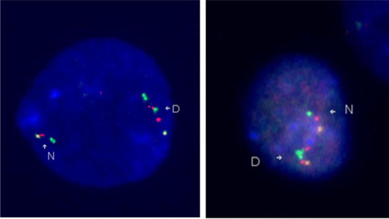
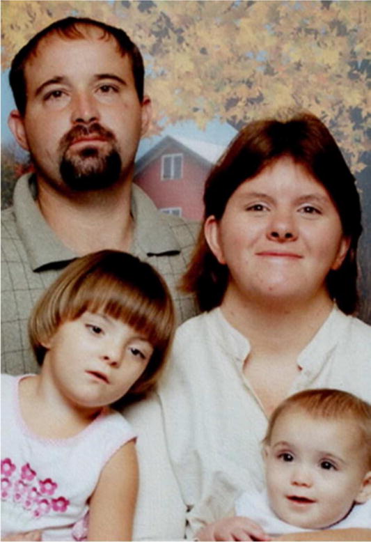
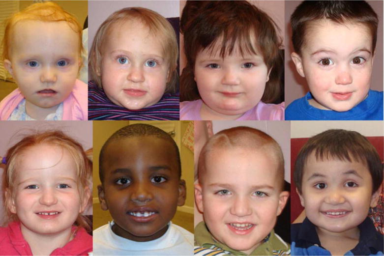
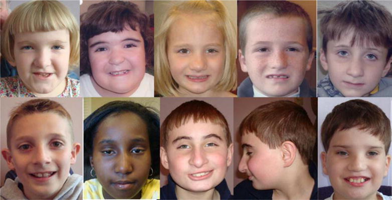
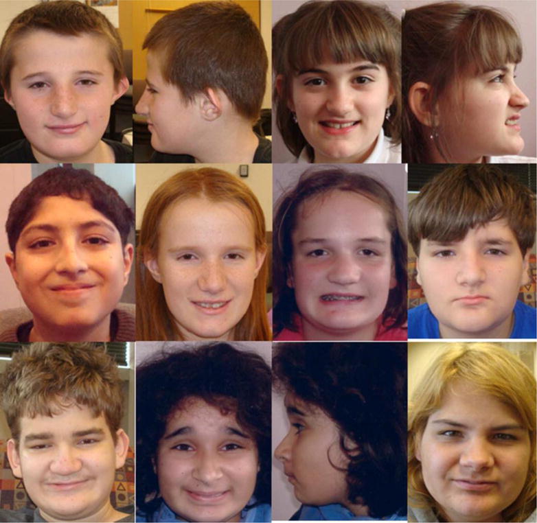
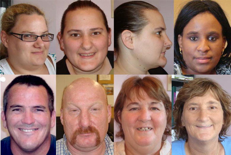
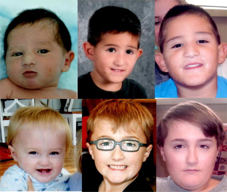
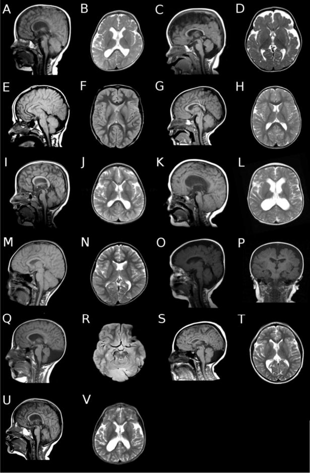
In order to describe the physical characteristics, medical complications, and natural history of classic 7q11.23 duplication syndrome [hereafter Dup7 (MIM 609757)], reciprocal duplication of the region deleted in Williams syndrome [hereafter WS (MIM 194050)], we systematically evaluated 53 individuals aged 1.25-21.25 years and 11 affected adult relatives identified in cascade testing. In this series, 27% of probands with Dup7 had an affected parent. Seven of the 26 de novo duplications that were examined for inversions were inverted; in all seven cases one of the parents had the common inversion polymorphism of the WS region. We documented the craniofacial features of Dup7: brachycephaly, broad forehead, straight eyebrows, broad nasal tip, low insertion of the columella, short philtrum, thin upper lip, minor ear anomalies, and facial asymmetry. Approximately 30% of newborns and 50% of older children and adults had macrocephaly. Abnormalities were noted on neurological examination in 88.7% of children, while 81.6% of MRI studies showed structural abnormalities such as decreased cerebral white matter volume, cerebellar vermis hypoplasia, and ventriculomegaly. Signs of cerebellar dysfunction were found in 62.3%, hypotonia in 58.5%, Developmental Coordination Disorder in 74.2%, and Speech Sound Disorder in 82.6%. Behavior problems included anxiety disorders, ADHD, and oppositional disorders. Medical problems included seizures, 19%; growth hormone deficiency, 9.4%; patent ductus arteriosus, 15%; aortic dilation, 46.2%; chronic constipation, 66%; and structural renal anomalies, 18%. We compare these results to the WS phenotype and offer initial recommendations for medical evaluation and surveillance of individuals who have Dup7.

Keywords: 7q11.23 duplication syndrome; Williams syndrome; anxiety; aortic dilation; cerebellar vermis hypoplasia; developmental coordination disorder; macrocephaly; psychopathology; speech sound disorder.


Figures

Figure 1
Figure 1
FISH images of a direct duplication and an inverted duplication of the WS region. The left image demonstrates a direct duplication. The N indicates the normal, non-duplicated, chromosome, with the probe order being yellow-red-green. The yellow is an anchor probe (RP11-815K3 (815)), and the red (BCL7 (BCL)) and green (RP5-1186P10 (1186)) probes are inside the WS region. The D indicates the duplicated chromosome. The probe order is yellow-red-green-red-green, or 815-BCL-1186-BCL-1186. Since the duplicated region, the extra red and green signals, is in the same orientation as the original region, the duplicated segment has been inserted in the normal orientation. The right image demonstrates an inverted duplication. The N indicates the normal chromosome and the D indicates the duplicated chromosome. The probe color order is yellow-red-green-green-red, or 815-BCL-1186-1186-BCL. Since the duplicated segment is in the opposite orientation, green-red, instead of the normal orientation, red-green, the duplicated segment has been inserted in the opposite direction, giving an inverted duplication.
Figure 2
Figure 2
Family with Dup7. The father, patient #43, transmitted the duplication to his daughters, patients #41 and 42. The mother of the children had a history of intellectual disability and died secondary to complications of a seizure disorder, but did not have Dup7. Patient #42 (the girl on the left) resembles her mother.
Figure 3
Figure 3
Facial features of Dup7 in young children. Top row left to right: 1.25 years, 1.5 years, 2.17 years, 2.5 years. Bottom row left to right: 3.9 years, 4.25 years, 4.25 years, 4.29 years.
Figure 4
Figure 4
Facial features of Dup7 in mid childhood. Top row left to right: 5.7 years, 5.91 years, 6.78 years, 7.09 years, 7.3 years. Bottom row left to right: 7.99 years, 9.75 years, 10 years (front and profile), 10.3 years.
Figure 5
Figure 5
Facial features of adolescents with Dup7. Top row left to right: 10.5 years (front and profile), 11 years (front and profile). Middle row left to right: 12.18 years, 13.25 years, 13.67 years, 13.99 years. Bottom row left to right: 13.99 years, 15.28 years (front and profile), 18.9 years.
Figure 6
Figure 6
Facial features of adults with Dup7. Top row left to right: 30 years, 31 years (front and profile), 33 years. Bottom row left to right: 39 years, 44 years, 49 years, 60 years.
Figure 7
Figure 7
Facial features over time. Top row: a boy at ages 1 day, 4 years, and 5 years. Bottom row: a boy at ages 6 months, 7 years, 11.6 years.
Figure 8
Figure 8
Representative brain MRI findings in individuals with Dup7. (A) Sagittal and (B) axial images of patient #1 showing thin corpus callosum and white matter, with mildly dilated lateral ventricles. (C) Sagittal and (D) axial images of patient #5 showing cerebellar vermis hypoplasia, thin corpus callosum and white matter, and increased cortical extra-axial space. (E) Sagittal and (F) axial images of patient #9 showing collapsed ventricles status post ventricular peritoneal shunting for hydrocephalus, and mildly increased extra-axial space in the posterior fossa. (G) Sagittal and (H) axial images of patient #12 showing normal brain morphology. (I) Sagittal and (J) axial images of patient #13 showing rounded corpus callosum, thin white matter, mild dilation of the lateral ventricles, and mild cerebellar vermis hypoplasia. (K) Sagittal and (L) axial images of patient #15 showing rounded corpus callosum, moderate dilation of the lateral ventricles, and thin white matter. There is also mild cerebellar tonsillar ectopia. (M) Sagittal and (N) axial images of patient #22 showing mild asymmetry of the lateral ventricles, probably due to mildly thin white matter, but otherwise normal brain morphology. (O) Sagittal and (P) coronal images of patient #23 showing increased extra axial spaces around both the cortex and in the posterior fossa. (Q) Sagittal and (R) axial images of patient #26, showing cerebellar vermis hypoplasia, and extra axial fluid collection in the posterior fossa. (S) Sagittal and (T) axial images of patient #29 showing mildly thin white matter, mildly dilated lateral ventricles, and cerebellar vermis hypoplasia with an extra axial fluid collection in the posterior fossa. (U) Sagittal and (V) axial images of patient #56 showing rounded corpus callosum, thin white matter, and moderately dilated lateral ventricles. The cerebellar vermis is mildly hypoplastic.

References

    1. Berg JS, Brunetti-Pierri N, Peters SU, Kang S-HL, Fong CT, Salamone J, Freedenberg D, Hannig VL, Prock LA, Miller DT, Raffalli P, Harris DJ, Erickson RP, Cunniff C, Clark GD, Blazo MA, Peiffer DA, Gunderson KL, Sahoo T, Patel A, Lupski JR, Beaudet AL, Cheung SW. Speech delay and autism spectrum behaviors are frequently associated with duplication of the 7q11.23 Williams-Beuren syndrome region. Genet Med. 2007;9:427–441. - PubMed
    1. Brown TA, DiNardo P, Barlow DH. Anxiety Disorder Interview Schedule Adult Version (ADIS-IV) San Antonio, TX: Graywind Publications; 1996.
    1. Collins RT, 2nd, Kaplan P, Somes GW, Rome JJ. Long-term outcomes of patients with cardiovascular abnormalities and Williams syndrome. Am J Cardiol. 2010;105:874–878. - PubMed
    1. Değerliyurt A, Ceylaner S, Ozdağ H. A 7q11.23 microduplication patient with cerebral palsy and facial dysmorphism. Genet Couns. 2012;23:263–267. - PubMed
    1. Depienne C, Heron D, Betancur C, Benyahia B, Trouillard O, Bouteiller D, Verloes A, Leguern E, Leboyer M, Brice A. Autism, language delay and mental retardation in a patient with 7q11 duplication. J Med Genet. 2007;44:452–458. - PMC - PubMed

Publication types

Supplementary concepts