

Predicting the Response of Breast Cancer to Neoadjuvant Therapy Using a Mechanically Coupled Reaction-Diffusion Model
- PMID: 26333809
- PMCID: PMC4651826
- DOI: 10.1158/0008-5472.CAN-14-2945
Predicting the Response of Breast Cancer to Neoadjuvant Therapy Using a Mechanically Coupled Reaction-Diffusion Model
Abstract
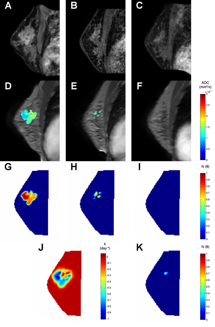
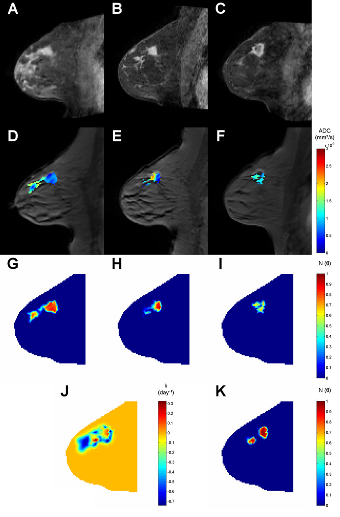
Although there are considerable data on the use of mathematical modeling to describe tumor growth and response to therapy, previous approaches are often not of the form that can be easily applied to clinical data to generate testable predictions in individual patients. Thus, there is a clear need to develop and apply clinically relevant oncologic models that are amenable to available patient data and yet retain the most salient features of response prediction. In this study we show how a biomechanical model of tumor growth can be initialized and constrained by serial patient-specific magnetic resonance imaging data, obtained at two time points early in the course of therapy (before initiation and following one cycle of therapy), to predict the response for individual patients with breast cancer undergoing neoadjuvant therapy. Using our mechanics coupled modeling approach, we are able to predict, after the first cycle of therapy, breast cancer patients that would eventually achieve a complete pathologic response and those who would not, with receiver operating characteristic area under the curve (AUC) of 0.87, sensitivity of 92%, and specificity of 84%. Our approach significantly outperformed the AUCs achieved by standard (i.e., not mechanically coupled) reaction-diffusion predictive modeling (0.75), simple analysis of the tumor cellularity estimated from imaging data (0.73), and the Response Evaluation Criteria in Solid Tumors (0.71). Thus, we show the potential for mathematical model prediction for use as a prognostic indicator of response to therapy. The work indicates the considerable promise of image-driven biophysical modeling for predictive frameworks within therapeutic applications.
©2015 American Association for Cancer Research.
Conflict of interest statement
Figures


Similar articles
-
Early Prediction of Breast Cancer Therapy Response using Multiresolution Fractal Analysis of DCE-MRI Parametric Maps.Tomography. 2019 Mar;5(1):90-98. doi: 10.18383/j.tom.2018.00046. Tomography. 2019. PMID: 30854446 Free PMC article.
-
Multiparametric magnetic resonance imaging for predicting pathological response after the first cycle of neoadjuvant chemotherapy in breast cancer.Invest Radiol. 2015 Apr;50(4):195-204. doi: 10.1097/RLI.0000000000000100. Invest Radiol. 2015. PMID: 25360603 Free PMC article.
-
Incorporating drug delivery into an imaging-driven, mechanics-coupled reaction diffusion model for predicting the response of breast cancer to neoadjuvant chemotherapy: theory and preliminary clinical results.Phys Med Biol. 2018 May 17;63(10):105015. doi: 10.1088/1361-6560/aac040. Phys Med Biol. 2018. PMID: 29697054 Free PMC article.
-
Meta-analysis of magnetic resonance imaging in detecting residual breast cancer after neoadjuvant therapy.J Natl Cancer Inst. 2013 Mar 6;105(5):321-33. doi: 10.1093/jnci/djs528. Epub 2013 Jan 7. J Natl Cancer Inst. 2013. PMID: 23297042 Review.
-
Quantitative magnetic resonance imaging and tumor forecasting of breast cancer patients in the community setting.Nat Protoc. 2021 Nov;16(11):5309-5338. doi: 10.1038/s41596-021-00617-y. Epub 2021 Sep 22. Nat Protoc. 2021. PMID: 34552262 Free PMC article. Review.
Cited by
-
Calibrating a Predictive Model of Tumor Growth and Angiogenesis with Quantitative MRI.Ann Biomed Eng. 2019 Jul;47(7):1539-1551. doi: 10.1007/s10439-019-02262-9. Epub 2019 Apr 8. Ann Biomed Eng. 2019. PMID: 30963385 Free PMC article.
-
Diffusion-weighted imaging in identifying breast cancer pathological response to neoadjuvant chemotherapy: A meta-analysis.Oncotarget. 2017 Dec 11;9(6):7088-7100. doi: 10.18632/oncotarget.23195. eCollection 2018 Jan 23. Oncotarget. 2017. PMID: 29467952 Free PMC article.
-
Computational reactive-diffusive modeling for stratification and prognosis determination of patients with breast cancer receiving Olaparib.Sci Rep. 2023 Jul 24;13(1):11951. doi: 10.1038/s41598-023-38760-z. Sci Rep. 2023. PMID: 37488154 Free PMC article. Clinical Trial.
-
The 2019 mathematical oncology roadmap.Phys Biol. 2019 Jun 19;16(4):041005. doi: 10.1088/1478-3975/ab1a09. Phys Biol. 2019. PMID: 30991381 Free PMC article. Review.
-
Current and Emerging Magnetic Resonance-Based Techniques for Breast Cancer.Front Med (Lausanne). 2020 May 12;7:175. doi: 10.3389/fmed.2020.00175. eCollection 2020. Front Med (Lausanne). 2020. PMID: 32478083 Free PMC article. Review.
References
-
- Martin I, Dozin B, Quarto R, Cancedda R, Beltrame F. Computer-based technique for cell aggregation analysis and cell aggregation in in vitro chondrogenesis. Cytometry. 1997;28:141–146. - PubMed
-
- Anderson AW, Xie J, Pizzonia J, Bronen RA, Spencer DD, Gore JC. Effects of cell volume fraction changes on apparent diffusion in human cells. Magn Reson Imaging. 2000;18:689–695. - PubMed
-
- Garg I, Miga MI. Preliminary investigation of the inhibitory effects of mechanical stress in tumor growth. SPIE Medical Imaging 2008: Visualization, Image-Guided Procedures, and Modeling Conference. 2008;6918
Publication types
MeSH terms
Grants and funding
- U01 CA142565/CA/NCI NIH HHS/United States
- 2R01NS049251/NS/NINDS NIH HHS/United States
- P30CA68485/CA/NCI NIH HHS/United States
- R01 CA138599/CA/NCI NIH HHS/United States
- R25CA092043/CA/NCI NIH HHS/United States
- 1U01CA142565/CA/NCI NIH HHS/United States
- R01 NS049251/NS/NINDS NIH HHS/United States
- 1P50 098131/PHS HHS/United States
- R25 CA092043/CA/NCI NIH HHS/United States
- P50 CA098131/CA/NCI NIH HHS/United States
- P30 CA068485/CA/NCI NIH HHS/United States
- R01CA138599/CA/NCI NIH HHS/United States
- U01CA174706/CA/NCI NIH HHS/United States
- U01 CA174706/CA/NCI NIH HHS/United States
LinkOut - more resources
Full Text Sources
Medical
