. 2015 Sep 2;7(303):303ra137.
doi: 10.1126/scitranslmed.aac4358.

ACVR1R206H receptor mutation causes fibrodysplasia ossificans progressiva by imparting responsiveness to activin A

Affiliations

ACVR1R206H receptor mutation causes fibrodysplasia ossificans progressiva by imparting responsiveness to activin A

Sarah J Hatsell et al. Sci Transl Med. .

Abstract

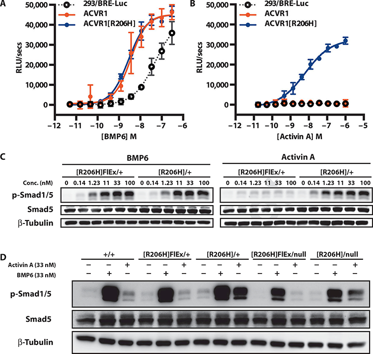
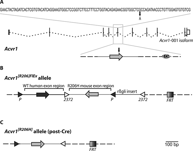
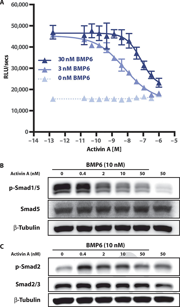
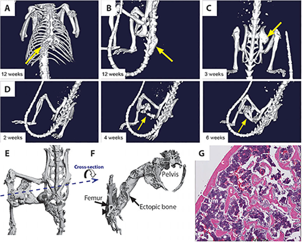
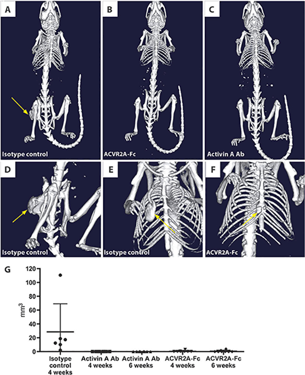
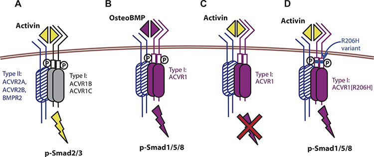
Fibrodysplasia ossificans progressiva (FOP) is a rare genetic disorder characterized by episodically exuberant heterotopic ossification (HO), whereby skeletal muscle is abnormally converted into misplaced, but histologically normal bone. This HO leads to progressive immobility with catastrophic consequences, including death by asphyxiation. FOP results from mutations in the intracellular domain of the type I BMP (bone morphogenetic protein) receptor ACVR1; the most common mutation alters arginine 206 to histidine (ACVR1(R206H)) and has been thought to drive inappropriate bone formation as a result of receptor hyperactivity. We unexpectedly found that this mutation rendered ACVR1 responsive to the activin family of ligands, which generally antagonize BMP signaling through ACVR1 but cannot normally induce bone formation. To test the implications of this finding in vivo, we engineered mice to carry the Acvr1(R206H) mutation. Because mice that constitutively express Acvr1[R206H] die perinatally, we generated a genetically humanized conditional-on knock-in model for this mutation. When Acvr1[R206H] expression was induced, mice developed HO resembling that of FOP; HO could also be triggered by activin A administration in this mouse model of FOP but not in wild-type controls. Finally, HO was blocked by broad-acting BMP blockers, as well as by a fully human antibody specific to activin A. Our results suggest that ACVR1(R206H) causes FOP by gaining responsiveness to the normally antagonistic ligand activin A, demonstrating that this ligand is necessary and sufficient for driving HO in a genetically accurate model of FOP; hence, our human antibody to activin A represents a potential therapeutic approach for FOP.


Conflict of interest statement

Competing interests: All authors except P.B.Y. are employees of Regeneron Pharmaceuticals Inc. and hold stock in the company. Regeneron has filed patent applications—US2014/0283158, which pertains to the mouse model, and US2015/0037339, which pertains to the use of anti-activin A neutralizing antibodies as therapy for FOP.

Figures

Fig. 1.
Fig. 1.. Acvr1 [R206H] signaling to Smad 1/5/8 is stimulated by activin A.
(A and B) Stable pools of HEK293/BRE-Luc reporter cells overexpressing human ACVR1 (solid red circles) or ACVR1[R206H] (solid blue circles) were treated overnight with BMP6 (A) or activin A (B), and activation of canonical BMP signaling was visualized using the Smad 1/5/8 reporter BRE-Luc. The response of the parental line is shown for comparison (293/BRE-Luc; open gray circles). (C) Mouse ES cells Acvr1[R206H]FlEx/+; Gt(ROSA26)SorCreERT2/+ and its post-Cre counterpart, Acvr1[R206H]FIEx/+; Gt(ROSA26)SorCreERT2/+/, were serum-starved for 2 hours and then treated with BMP6 or activin A for 1 hour. (D) Comparison of BMP6- or activin A-stimulated signaling in Acvr1+/+ (+/+), Acvr1[R206H]FlEx/+ ([R206H]FIEx/+), Acvr1[R206H]/+ ([R206H]/+), Acvr1[R206H]FlEx/nuII ([R206H]FIEx/null), and Acvr1[R206H1/nuII ([R206H]/null) ES cells. All lines also carry Gt(ROSA26)SorCreERT2/+. Response to ligands was visualized by Western blotting for phospho-Smad1/5. Total Smad5 and β-tubulin (loading control) are shown for comparison. RLU, relative light unit. RLUs are background-subtracted; error bars represent SD from samples run in triplicate.
Fig. 2.
Fig. 2.. A conditional-on knock-in allele of ACVR1R206H.
(A) Sequence of exon 5 along with the exon-intron structure of Acvr1 isoform 001 (adapted from www.Ensembl.org). The G-to-A missense mutation that results in the R206H amino acid change is marked. LOAp marks the small intronic region deleted for genotyping purposes (see Materials and Methods). (B) Structure of the Acvr1[R206H]FlEx allele. Mouse Acvr1 exon 5 and associated intronic sequence (R206H mouse exon region) have been placed in the antisense strand of Acvr1 within introns 5 to 6 and have been altered to encode R206H (*). The corresponding human exon and associated intronic sequence have been inserted in the sense strand to replace its corresponding mouse counterpart [wild-type (WT) human exon region]. These two regions have been flanked by FlEx arrays, arranged in a manner such that, upon action of Cre, the introduced human sequence will be deleted and the R206H mouse exon region will be brought to the sense strand, thereby giving rise to an Acvr1[R206H] allele. To ensure efficient action by Cre, the loxP and lox2372 sites of the 3′FlEx array have been separated by a small insert derived from rabbit hemoglobin β intron 2 (rβgli insert). An FRT site also remains after removal of the drug selection cassette (not shown). (C) Structure of the Acvr1[R206H] allele. Mouse Acvr1 exon 5 and associated intronic sequence have been placed in the sense strand of Acvr1.Note that the only extraneous elements that remain after action by Cre are two lox sites, an FRT site, and the small deletion of the region initially marked as LOAp in (A). Light gray, mouse sequences; dark gray, human sequences; dotted line, unaltered mouse genomic sequence in the Acvr1 locus.
Fig. 3.
Fig. 3.. Activin A inhibits BMP6 signaling via ACVR1.
(A) The HEK293/BRE-Luc reporter line expressing WT ACVR1 was stimulated with 0,3, or 30 nM BMP6 and varying amounts (dose curve) of activin A. Error bars represent SD from samples run in triplicate. Lowest concentration on dose response represents no addition of activin A. (B and C) Competition assays were performed in WT ES cells to examine whether the activin competition for BMP6 signaling could result from an increase in Smad2 phosphorylation. Samples were analyzed for phospho-Smad1/5 (B) and phospho-Smad 2 (C) by Western blotting.
Fig. 4.
Fig. 4.. Heterotopic boneformation in Acvr1[R206H]FlEx/+; Gt(ROSA26)SorCreERT2/+ mice after tamoxifen treatment.
(A to D) Representative examples of in vivo μCT images from Acvr1[R206H]FlEx/+; Gt(ROSA26)SorCreERT2/+ mice 2 to 12 weeks after tamoxifen administration. Ectopic bone growth was found at a number of different sites including adjacent to the existing skeleton in the (A) sternum, (B) caudal vertebrae, and (C) hip joint. (D) Ectopic bone growth formed between 2 and 4 weeks after tamoxifen injection and could occur distal to the existing skeleton. (E) Ex vivo μCT image of an ectopic bone lesion from the dorsal side showing bridging from the femur to the pelvis. (F) A transverse view through the ectopic bone shows that the newly formed bone has both cortical and trabecular like structures. At the region of intersection of the ectopic bone and the endogenous skeleton (arrow), there is evidence of remodeling of the cortical bone, but no evidence of bone marrow sharing. (G) Hematoxylin and eosin (H&E)-stained histological sections of ectopic bone lesion demonstrating cortical (c) and trabecular (t) bone-like structures and bone marrow (bm).
Fig. 5.
Fig. 5.. ACVR2A-Fc and activin A antibodies prevent HO in Acvr1[R206H]FlEx/+; Gt(ROSA26)SorCreERT2/+ mice.
Representative in vivo μCT images of mice at 3 weeks after initiation of the FOP model. (A) HO formation in a mouse treated with isotype control antibody (25 mg/kg, twice weekly). (B) No evidence of HO formation in a mouse treated with ACVR2A-Fc (10 mg/kg, twice weekly). (C) No evidence of HO in mice treated with activin A antibody (Ab) (25 mg/kg). (D and E) Large HO lesions at various locations including the hip joint and rib cage in mice treated with an isotype control antibody. (F) A small HO lesion found in two of the mice treated with ACVR2A-Fc. (G) Quantification of the total volume of HO lesions in each mouse at 4 weeks (isotype control, n = 6; activin A antibody, n = 8; ACVR2B-Fc, n = 8) and 6 weeks (activin A antibody; ACVR2B-Fc) after initiation of the model.
Fig. 6.
Fig. 6.. An intracellular domain amino acid change (R206H) transforms ACVR1 into an activin-responsive receptor.
(A) Activin signals via the type I receptors ACVR1B/1C and Smad2/3 phosphorylation but shares type II receptors (ACVR2A, ACVR2B, and BMPR2) with BMPs. (B) ACVR1, together with the type II receptors, recognizes BMPs and stimulates phosphorylation of Smad1/5/8. (C) ACVR1, together with the type II receptors, binds activin, but the resulting complex does not stimulate phosphorylation of Smad1/5/8; instead, activin acts as an inhibitor of canonical BMP-mediated signaling through ACVR1. (D) The R206H variant of the ACVR1 receptor responds to activin, inducing Smad1/5/8 phosphorylation, just like a BMP, effectively converting the ACVR2-ACVR1-activin complex from a dead end complex into a signaling complex. The R206H variant does not lose its ability to respond to canonical BMPs. The ACVR2-ACVR1-activin complex is shown here to contain a heterodimer of ACVR1-ACVR1[R206H]; however, this is not an obligate arrangement because a homodimer of ACVR1[R206H] is also capable of transducing the signal.

Comment in

References

    1. Hüning I, Gillessen-Kaesbach G, Fibrodysplasia ossificans progressiva: Clinical course, genetic mutations and genotype-phenotype correlation. Mol. Syndromol 5, 201–211 (2014). - PMC - PubMed
    1. Shore EM, Xu M, Feldman GJ, Fenstermacher DA, Cho TJ, Choi IH, Connor JM, Delai P, Glaser DL, LeMerrer M, Morhart R, Rogers JG, Smith R, Triffitt JT, Urtizberea JA, Zasloff M, Brown MA, Kaplan FS,A recurrent mutation in the BMP type I receptor ACVR1 causes inherited and sporadic fibrodysplasia ossificans progressiva. Nat. Genet 38, 525–527 (2006). - PubMed
    1. Groppe JC, Shore EM, Kaplan FS, Functional modeling of the ACVR1 (R206H) mutation in FOP. Clin. Orthop. Relat. Res 462, 87–92 (2007). - PubMed
    1. Groppe JC, Wu J, Shore EM, Kaplan FS, In vitro analyses of the dysregulated R206H ALK2 kinase-FKBP12 interaction associated with heterotopic ossification in FOP. Cells Tissues Organs 194, 291–295 (2011). - PMC - PubMed
    1. Chaikuad A, Alfano I, Kerr G, Sanvitale CE, Boergermann JH, Triffitt JT, von Delft F, Knapp S, Knaus P, Bullock AN, Structure of the bone morphogenetic protein receptor ALK2 and implications for fibrodysplasia ossificans progressiva. J. Biol. Chem 287, 36990–36998 (2012). - PMC - PubMed