. 2015 Dec;114(6):3097-110.
doi: 10.1152/jn.00618.2015. Epub 2015 Sep 2.

Processing time of addition or withdrawal of single or combined balance-stabilizing haptic and visual information

Affiliations

Processing time of addition or withdrawal of single or combined balance-stabilizing haptic and visual information

Jean-Louis Honeine et al. J Neurophysiol. 2015 Dec.

Abstract

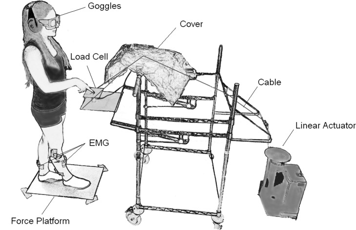
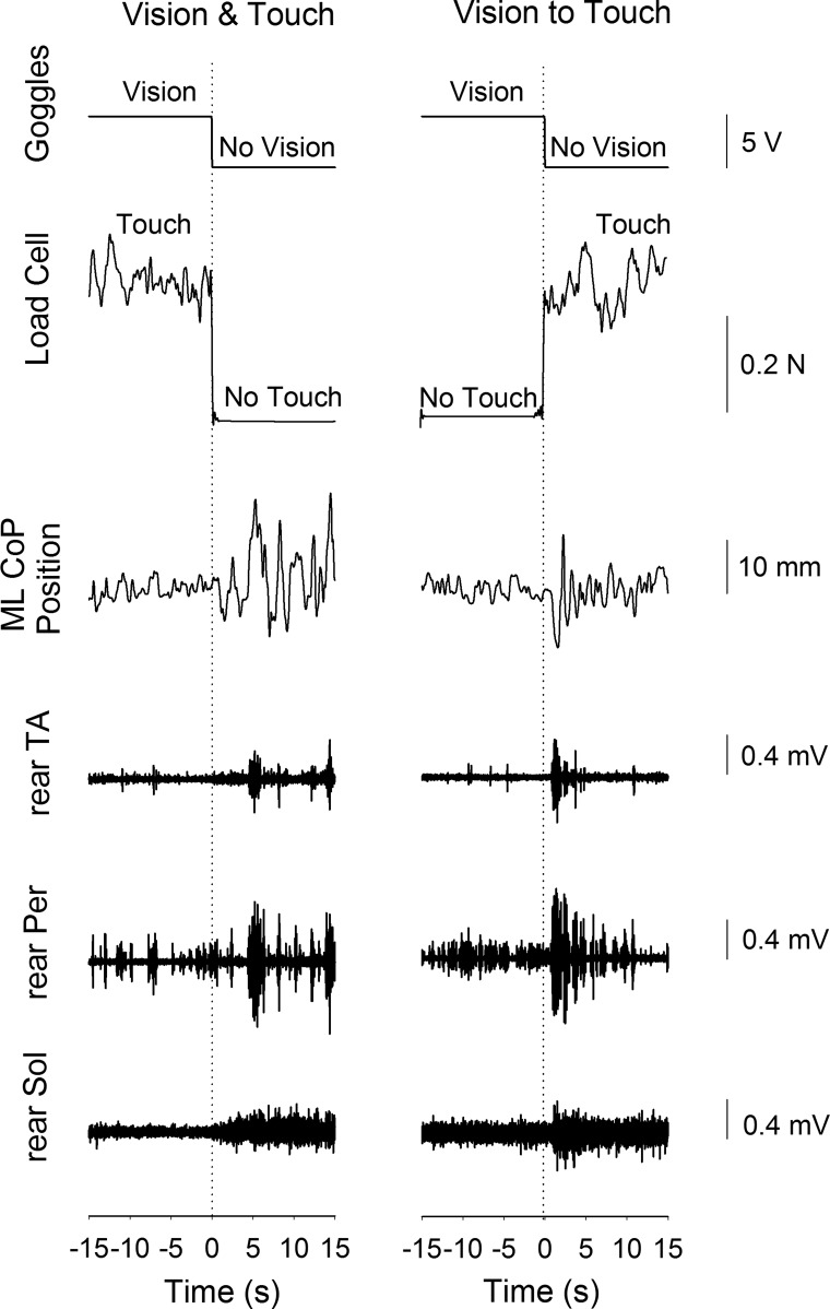
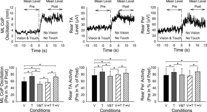
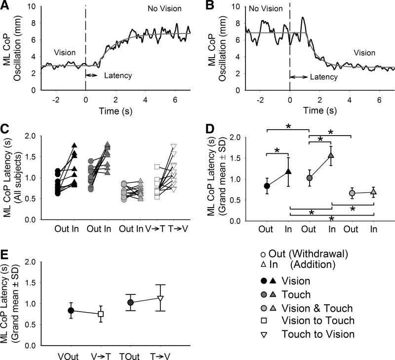
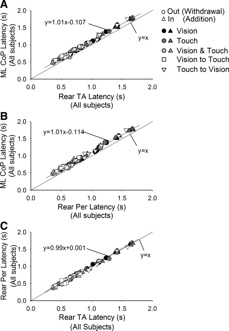
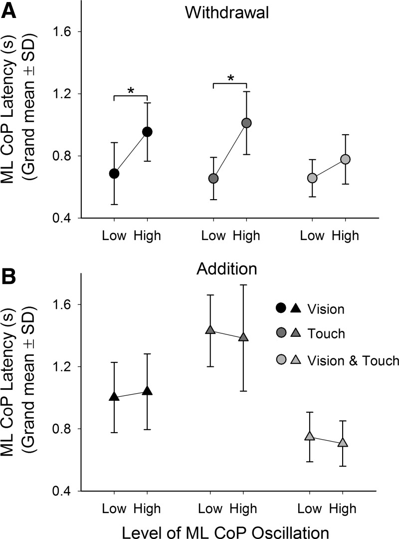
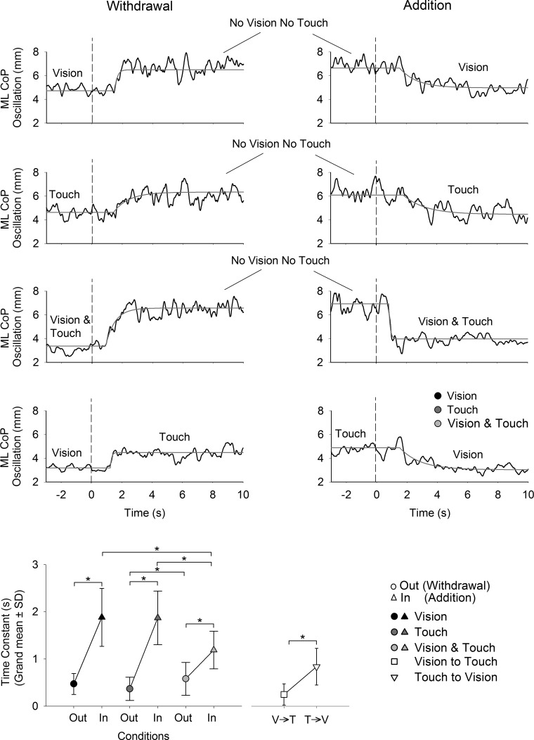
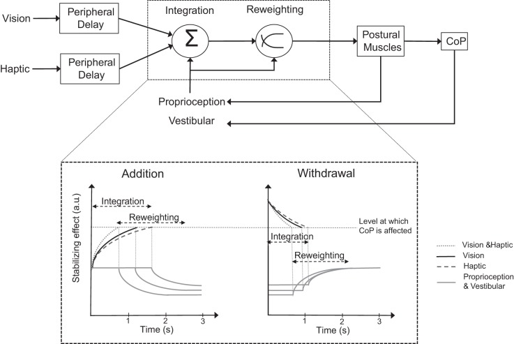
We investigated the integration time of haptic and visual input and their interaction during stance stabilization. Eleven subjects performed four tandem-stance conditions (60 trials each). Vision, touch, and both vision and touch were added and withdrawn. Furthermore, vision was replaced with touch and vice versa. Body sway, tibialis anterior, and peroneus longus activity were measured. Following addition or withdrawal of vision or touch, an integration time period elapsed before the earliest changes in sway were observed. Thereafter, sway varied exponentially to a new steady-state while reweighting occurred. Latencies of sway changes on sensory addition ranged from 0.6 to 1.5 s across subjects, consistently longer for touch than vision, and were regularly preceded by changes in muscle activity. Addition of vision and touch simultaneously shortened the latencies with respect to vision or touch separately, suggesting cooperation between sensory modalities. Latencies following withdrawal of vision or touch or both simultaneously were shorter than following addition. When vision was replaced with touch or vice versa, adding one modality did not interfere with the effect of withdrawal of the other, suggesting that integration of withdrawal and addition were performed in parallel. The time course of the reweighting process to reach the new steady-state was also shorter on withdrawal than addition. The effects of different sensory inputs on posture stabilization illustrate the operation of a time-consuming, possibly supraspinal process that integrates and fuses modalities for accurate balance control. This study also shows the facilitatory interaction of visual and haptic inputs in integration and reweighting of stance-stabilizing inputs.

Keywords: haptic; sensory integration; sensory reweighting; standing; vision.


Figures

Fig. 1.
Fig. 1.
Experimental setup. This figure shows a subject standing in the tandem posture on the force platform over which the contour of the feet has been marked. The subject's state of vision was controlled via electronic goggles. A pulley system activated by a linear actuator pulling on a transparent cord (colored black here) allowed the experimenter to lower or raise a load cell, on which a wooden board (15 × 12 cm) was rigidly attached, to produce or withdraw haptic input passively with the index finger. The cord was hidden from the field of view of the subject by covering it with a tissue. Earmuffs were worn at all time to prevent the subjects from hearing any acoustic cues during the experiment. EMG, electromyogram.
Fig. 2.
Fig. 2.
Raw recorded signals. This figure illustrates the traces obtained during 1 trial of the Vision & Touch condition (left) and 1 trial of the Vision to Touch condition (right). From top to bottom are the transistor-transistor logic (TTL) signal used to control the goggles, the force measured by the load cell, the mediolateral (ML) position of the center of pressure (CoP), and the activity of tibialis anterior (TA), peroneus longus (Per), and soleus (Sol) of the rear leg, respectively. After a short period following the change in sensory state, ML CoP oscillation and muscle activity increased when vision and touch were withdrawn simultaneously (left) and when vision was replaced with touch (right).
Fig. 3.
Fig. 3.
Mean levels of CoP oscillation and muscle activity. Top panels show the mean levels (n = 60 trials) of ML CoP oscillation and rear TA and Per (from left to right) of 1 subject during Vision & Touch condition. The mean values were calculated in the time windows indicated by the arrows above the curves. The mean levels in the presence of stabilizing input (Mean Level Pre) were then calculated in percentage of the maximum levels in the absence of vision and touch (Mean Level Post). The bars of the bottom panels show the grand mean plus SD of the levels of CoP oscillation and rear TA and Per activity when vision “alone” (V), touch “alone” (T), vision and touch (V&T), vision to touch (V→T), and touch to vision (T→V) were present. The mean levels of CoP oscillation and muscle activity were reduced in the presence of sensory input. The lowest CoP oscillations were obtained in V&T, and the highest in T. The mean levels of CoP oscillation of V and T conditions are comparable with those obtained during the V→T and T→V conditions, respectively. The asterisks indicate significant differences.
Fig. 4.
Fig. 4.
Latencies of CoP oscillation following the change in sensory state. The upper panels show the mean curves of CoP oscillation of a representative subject during withdrawal (A) and addition (B) of V (black; at t = 0) and the fitted-exponential function (gray). The segments between the arrowheads under the curves indicate the latencies. In C, the latencies calculated for the CoP oscillation of each subject in all 4 conditions are presented. The grand mean and SD of the latencies in sway for the V, T, and V&T conditions are provided in D. In the labels of the abscissae of C and D, “Out” is used to indicate withdrawal and “In” addition (the symbol key is at the bottom right). The latencies were shorter on withdrawal than addition. They were longest following addition of T and shortest following addition of V&T. In E, the grand mean and SD of the latencies following withdrawal of V, withdrawal of T, and the replacement of V with T and vice versa are provided. The asterisks indicate significant differences.
Fig. 5.
Fig. 5.
Latencies of change in muscle activity following change in sensory state. Latencies of CoP oscillation levels are plotted against the latencies of rear TA (A) and rear Per (B) EMG for the different conditions (symbols as in Fig. 4), and the best-fit line is drawn. The identity line (y = x) is also indicated. Changes in muscle activity always preceded those of sway by ∼110 ms. In C, latencies of rear Per are plotted against those of rear TA. The data points sit on the identity line indicating equal mean latency between muscles for each condition.
Fig. 6.
Fig. 6.
Latency of change in CoP oscillation as a function of the oscillation level before sensory shift. All trials performed by each subject have been divided into 2 sets, 1 composed of low-level oscillation trials, the other of high-level oscillation trials. The grand means and SD of the latencies of each set in each condition (V, black; T, dark gray; V&T, light gray) are provided during withdrawal (A) and addition (B). Following withdrawal, the latencies observed in each condition were longer in the high-oscillation level trials. The same effect did not occur for addition. The asterisks indicate significant differences.
Fig. 7.
Fig. 7.
Time constant of the recovery to steady-state following the sensory shift. Upper panels show the mean curves of the CoP oscillation (black) and the fitted exponential function (gray) in all conditions. The grand mean and SD for the time constant calculated for all subjects in each condition are provided in the bottom graph. The times constants were generally shorter on withdrawal than addition of V and/or T. The time constants were also shorter following addition of V and T simultaneously with respect to the addition of V alone and T alone. The asterisks indicate significant differences.
Fig. 8.
Fig. 8.
Simple scheme of the sensorimotor integration and reweighting processes following addition or withdrawal of V and T during standing. Vestibular, proprioceptive, visual, and haptic (T) information are continuously detected and passed to the brain (top). The different modalities converge at an integrator, which incorporates the different stimuli and transforms them into information for maintaining balance. Following sudden shift in visual and/or haptic state, a variable integration time period occurs before any change in sway level is observed (bottom). Following addition of V (solid black), T (dashed), or both simultaneously (dotted), the integration process reaches the threshold (horizontal dashed line) at which the brain modifies the postural muscle activity at different times according to modality. When both V and T are added, the integration time is the shortest. In the case of withdrawal, the “disintegration” process is faster than addition, being the shortest when both V and T are removed. Once the threshold is reached following addition, CoP oscillation starts decreasing exponentially, reflecting the upweighting of the new stabilizing information and the downweighting of the proprioceptive and vestibular inflow (solid gray). Following withdrawal, the remaining proprioceptive and vestibular inputs are upweighted. a.u., Arbitrary units.

Similar articles

Cited by

References

    1. Assländer L, Hettich G, Mergner T. Visual contribution to human standing balance during support surface tilts. Hum Mov Sci 41: 147–164, 2015. - PMC - PubMed
    1. Assländer L, Peterka RJ. Sensory reweighting dynamics in human postural control. J Neurophysiol 111: 1852–1864, 2014. - PMC - PubMed
    1. Azañón E, Longo MR, Soto-Faraco S, Haggard P. The posterior parietal cortex remaps touch into external space. Curr Biol 20: 1304–1309, 2010. - PubMed
    1. Baudry S, Duchateau J. Independent modulation of corticospinal and group I afferents pathways during upright standing. Neuroscience 275: 162–169, 2014. - PubMed
    1. Baudry S, Penzer F, Duchateau J. Vision and proprioception do not influence the excitability of the corticomotoneuronal pathway during upright standing in young and elderly adults. Neuroscience 268: 247–254, 2014. - PubMed

Publication types

LinkOut - more resources