. 2015 Sep 8;6(26):21840-52.
doi: 10.18632/oncotarget.4808.

Molecular dissection of HBV evasion from restriction factor tetherin: A new perspective for antiviral cell therapy

Affiliations

Molecular dissection of HBV evasion from restriction factor tetherin: A new perspective for antiviral cell therapy

Kei Miyakawa et al. Oncotarget. .

Abstract

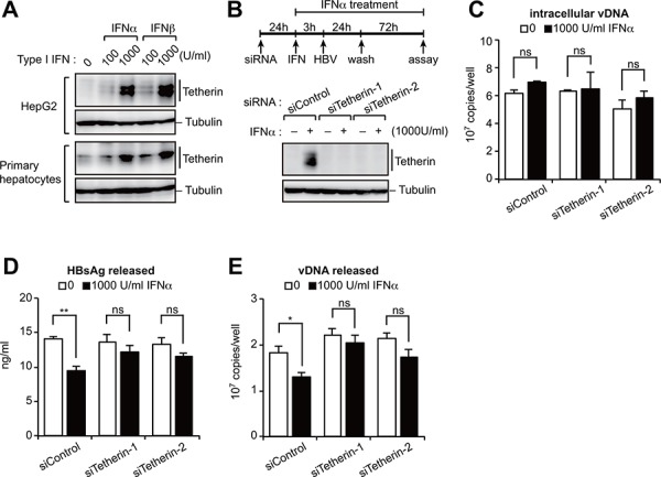
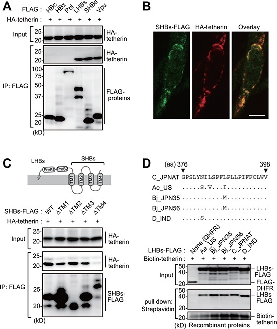
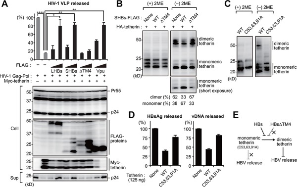
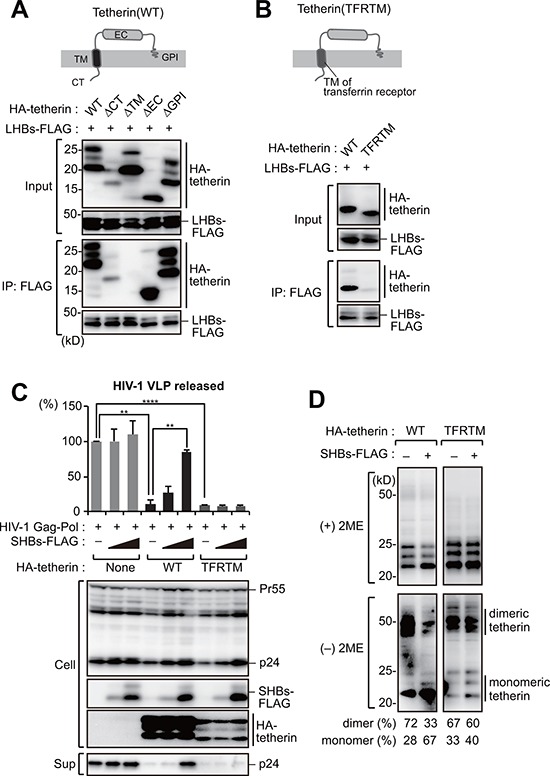
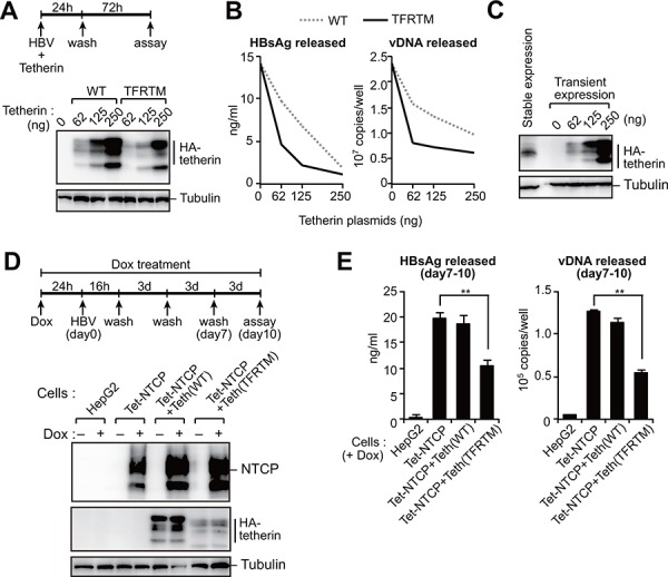
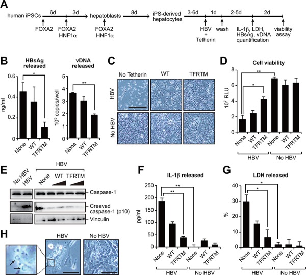
Viruses have evolved various strategies to escape from the innate cellular mechanisms inhibiting viral replication and spread. Extensive evidence has highlighted the ineffectiveness of interferon (IFN) therapy against chronic hepatitis B virus (HBV) infection, implying the existence of mechanisms by which HBV evades IFN-induced antiviral responses. In our current study, we demonstrate that HBV surface protein (HBs) plays a crucial role in counteracting the IFN-induced antiviral response mediated by tetherin (also known as BST-2). The type I IFN treatment of HBV-producing cells marginally but significantly inhibited the release of HBsAg and viral DNA, but this release was recovered by the knockdown of tetherin. HBs can interact with tetherin via its fourth transmembrane domain thereby inhibiting its dimerization and antiviral activity. The expression of a tetherin mutant devoid of the HBs-binding domain promoted a prominent restriction of HBV particle production that eventually resulted in the alleviation of caspase-1-mediated cytotoxicity and interleukin-1β secretion in induced pluripotent stem cell (iPSC)-derived hepatocytes. Our current results thus reveal a previously undescribed molecular link between HBV and tetherin during the course of an IFN-induced antiviral response. In addition, strategies to augment the antiviral activity of tetherin by impeding tetherin-HBs interactions may be viable as a therapeutic intervention against HBV.

Keywords: Immune response; Immunity; Immunology and Microbiology Section; hepatic injury; pyroptosis.


Conflict of interest statement

CONFLICTS OF INTEREST

No, there is no conflict of interest.

Figures

Figure 1
Figure 1. Type I IFN-induced tetherin weakly represses HBV release
A. Immunoblotting analysis of HepG2 cells (top) and primary hepatocytes (bottom) treated with either IFNα or IFNβ (100 or 1,000 U/ml) for 24 h before harvesting. B–E. HepG2 cells were transduced with siRNA targeting tetherin (siTetherin-1 or 2) or control siRNA, and were treated with IFNα, then transfected with an HBV molecular clone (pUC19-C_JPNAT) 24 h later. One day after transfection, the cells were washed and treated with IFNα for three days. The expression of tetherin and tubulin in cells was detected by immunoblotting (B). The amounts of viral DNA (vDNA) in cells (C) and in the culture supernatants (E) were measured by real-time PCR. The amounts of HBsAg in the culture supernatants were measured by ELISA (D). ns, not significant; *P < 0.05; **P < 0.01.
Figure 2
Figure 2. HBs binds tetherin via its fourth transmembrane domain
A. HEK293 cells were cotransfected with plasmids encoding HA-tetherin and the indicated FLAG-tagged HBV proteins. Cell lysates were immunoprecipitated with anti-FLAG antibody and then analyzed by immunoblotting with either anti-HA or anti-FLAG antibody. Vpu (tetherin-interacting HIV protein) was used as a positive control. B. HepG2 cells were transfected with plasmids encoding SHBs-FLAG and HA-tetherin. After 24 h, the cells were fixed, permeabilized, and stained with anti-FLAG (green) and anti-HA (red), followed by confocal microscopic analysis. Scale bar, 10 μm. C. Schematic representation of the domain structure of HBs (top). HEK293 cells were transfected with WT SHBs-FLAG or its transmembrane domain-deficient mutants (ΔTM) together with HA-tetherin. Cell lysates were immunoprecipitated with anti-FLAG antibody, and the bound proteins were analyzed by immunoblotting with either anti-HA or anti-FLAG antibody (bottom). D. Alignment of the HBs TM4 sequence with the indicated HBV variants (top). Recombinant FLAG-tagged LHBs derived from the indicated HBV genotypes were mixed with recombinant biotinylated tetherin and then processed for the in vitro pull-down analysis with streptavidin sepharose beads. Captured proteins were analyzed by immunoblotting with either anti-FLAG antibody or horseradish peroxidase-conjugated streptavidin (bottom). DHFR (dihydrofolate reductase) was used as a negative control.
Figure 3
Figure 3. HBs inhibits the dimerization of tetherin to counteract its antiviral activity
A. HIV-1 VLP release assay was performed in HepG2 cells transfected with the indicated vectors encoding HBs proteins (LHBs, SHBs and SHBsΔTM4) or Vpu (tetherin antagonist of HIV-1). At four days after transfection, the levels of p24 capsid protein in the culture supernatants were measured by ELISA (top). Blots below the bar graph show the detection of HIV-1 Gag (precursor Pr55 and p24 capsid), FLAG-tagged viral proteins and Myc-tetherin by immunoblotting (bottom). *P < 0.05; **P < 0.01; ****P < 0.0001. B, C. HEK293 cells were transfected with the vectors encoding HA-tetherin (100 ng) with or without SHBs expression plasmids (1 μg). Cell lysates were subjected to SDS-PAGE in the absence or presence of 2-mercaptoethanol (2ME), and analyzed by immunoblotting with anti-HA antibody. The numerical values below the blot indicate the amounts of dimer or monomer bands determined by densitometry. D. HepG2 cells were cotransfected with pUC19-C_JPNAT (2.5 μg) and vectors encoding WT or C53,63,91A tetherin (125 ng). At four days after transfection, the amounts of HBsAg and vDNA in the culture supernatants were measured. Note that 125 ng of WT tetherin can inhibit HBV release (see Figure 5B). E. Predicted model for the HBs-mediated counteraction of tetherin.
Figure 4
Figure 4. The transmembrane domain of tetherin is responsible for HBs binding
A. Schematic representation of the domain structure of tetherin (top). HEK293 cells were transfected with SHBs-FLAG and cotransfected with WT tetherin or its domain mutants (CT: cytoplasmic tail; TM: transmembrane; EC: extracellular domain, GPI: glycosylphosphatidylinositol anchor). Cell lysates were then immunoprecipitated with anti-FLAG antibody, and the bound proteins were analyzed by immunoblotting with either anti-HA or anti-FLAG antibody (bottom). B. Schematic representation of a substitution mutant of tetherin harboring a TM domain derived from transferrin receptor (TFRTM) (top). WT tetherin and its TFRTM mutant were subjected to the immunoprecipitation analysis as in (A) (bottom). C. HIV-1 VLP release assay in HepG2 cells transduced with WT tetherin and its TFRTM mutant together with the indicated vectors, as in Figure 3A. **P < 0.01; ***P < 0.001; ****P < 0.0001. D. Detection of tetherin dimerization in HEK293 cells transfected with the indicated vectors encoding HBs and HA-tetherin, as in Figure 3B. The numerical values below the blot indicate the amounts of dimer or monomer bands determined by densitometry.
Figure 5
Figure 5. HBs-resistant chimeric tetherin efficiently restricts HBV release
A, B. HepG2 cells were cotransfected with pUC19-C_JPNAT (2.5 μg) and the indicated amounts of expression vector encoding WT or TFRTM tetherin. Four days after transfection, the indicated protein expression in cells was detected by immunoblotting (A). The HBsAg or vDNA levels in the culture supernatants were measured by ELISA or real-time PCR, respectively (B). Data are representative of three experiments. C. Comparison of tetherin expression in stably- and transiently-expressing HepG2 cells. D, E. Indicated cells were infected with HBV in the presence or absence of Dox. At 11 days after infection, the indicated protein expression in cells was detected by immunoblotting (C). The amounts of HBsAg and vDNA in the culture supernatants were measured by ELISA and real-time PCR, respectively (D). **P < 0.01.
Figure 6
Figure 6. Transduction of TFRTM tetherin inhibits virus-induced cytotoxicity in iPSC-derived hepatocytes
A, B. Hepatocyte-like cells were differentiated from human induced pluripotent stem cells (iPSCs) according to a previously described method [42] (A). iPSC-derived hepatocytes were cotransfected with pUC19-C_JPNAT and vectors encoding WT or TFRTM tetherin. Four days after transfection, the amounts of HBsAg and vDNA in the culture supernatants were measured using ELISA and real-time PCR, respectively (B). *P < 0.05; **P < 0.01. C, D. Microscopic images (C) and cell viability (D) of indicated hepatocytes at 7 days after transfection. Scale bar, 200 μm. *P < 0.05; **P < 0.01. E–G. Detection of cleaved caspase-1 in cell lysates (E) and secreted IL-1β (F) and LDH (G) in the culture supernatants of indicated hepatocytes at 4 days after transfection. *P < 0.05; **P < 0.01. H. Plasma membrane raptures (black arrow) were mainly observed in HBV-transduced hepatocytes (at 3 days after transfection). Scale bar, 50 μm.

Similar articles

Cited by

References

    1. Ivashkiv LB, Donlin LT. Regulation of type I interferon responses. Nature reviews Immunology. 2014;14:36–49. - PMC - PubMed
    1. Goubau D, Deddouche S, Reis e Sousa C. Cytosolic sensing of viruses. Immunity. 2013;38:855–869. - PMC - PubMed
    1. Duggal NK, Emerman M. Evolutionary conflicts between viruses and restriction factors shape immunity. Nature reviews Immunology. 2012;12:687–695. - PMC - PubMed
    1. Dienstag JL. Hepatitis B virus infection. The New England journal of medicine. 2008;359:1486–1500. - PubMed
    1. Neil SJ. The antiviral activities of tetherin. Current topics in microbiology and immunology. 2013;371:67–104. - PubMed

Publication types

MeSH terms

LinkOut - more resources