. 2015 Sep 3:5:13734.
doi: 10.1038/srep13734.

CRISPR/Cas9 nickase-mediated disruption of hepatitis B virus open reading frame S and X

Affiliations

CRISPR/Cas9 nickase-mediated disruption of hepatitis B virus open reading frame S and X

Madina Karimova et al. Sci Rep. .

Abstract

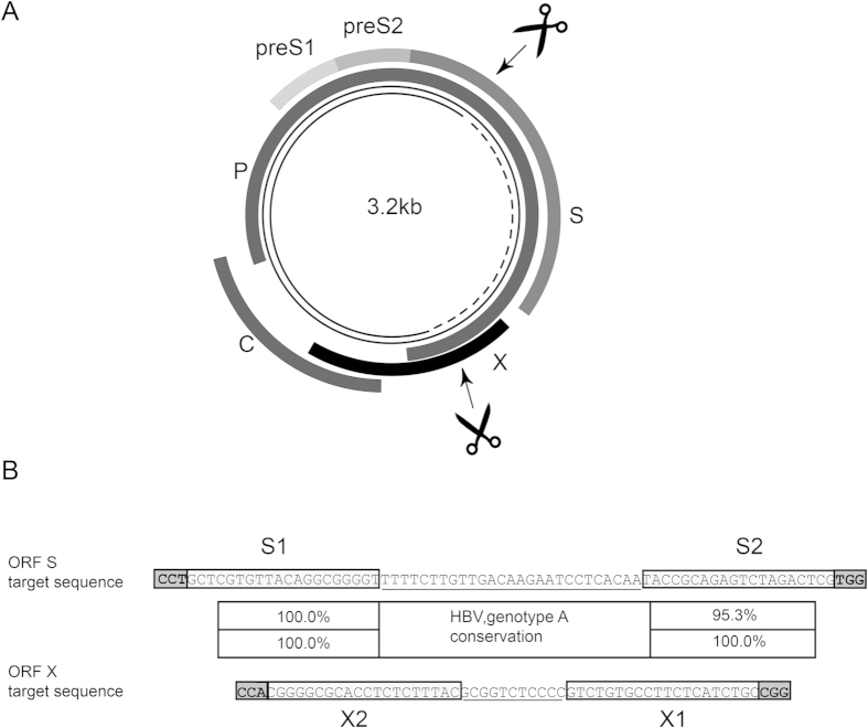
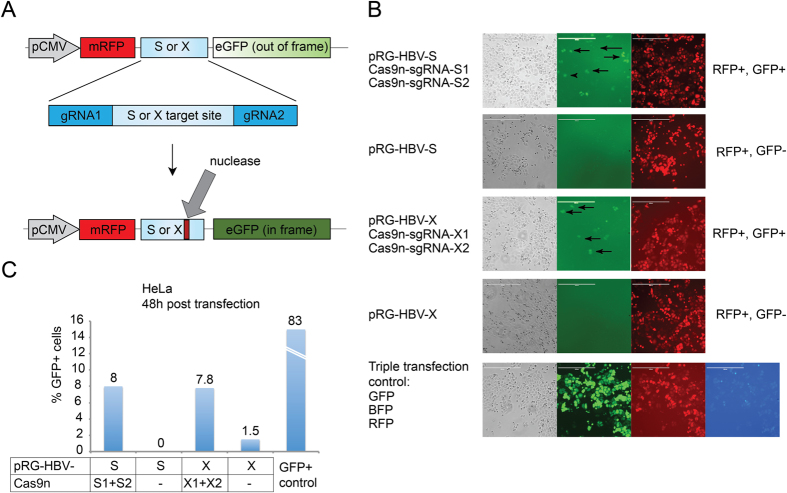
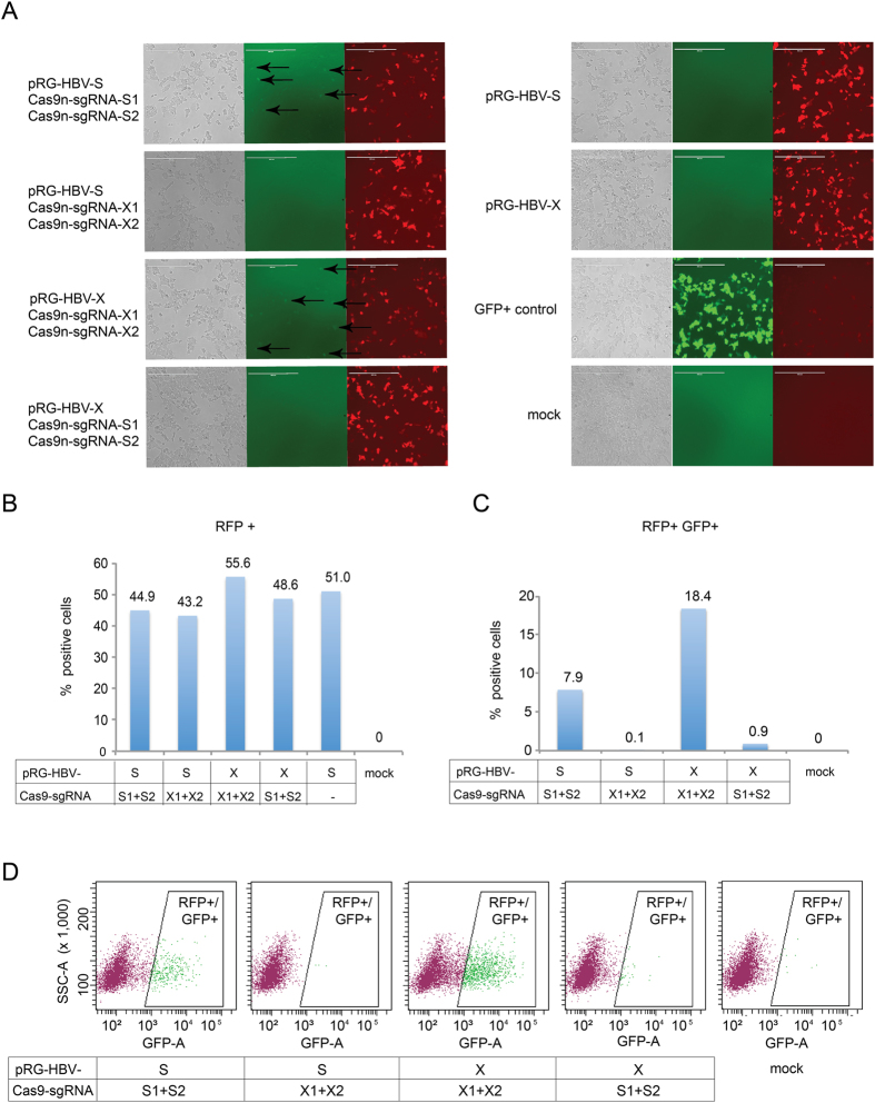
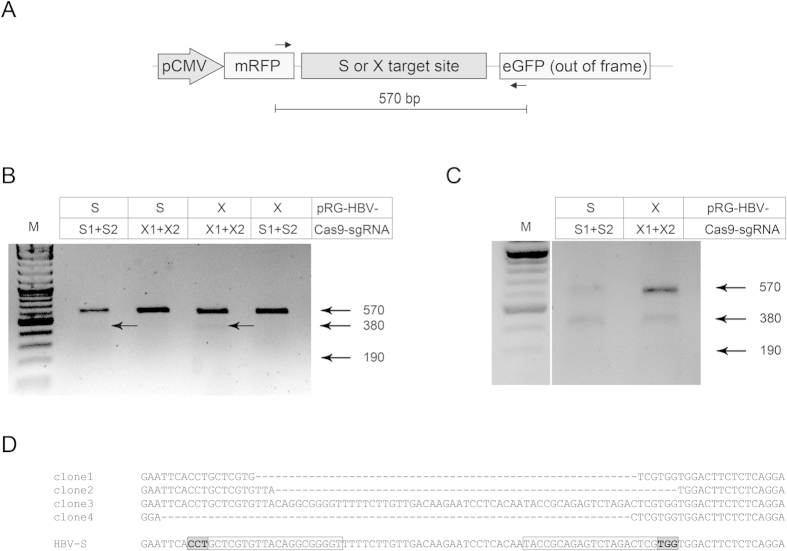
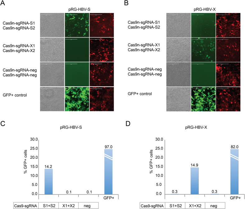
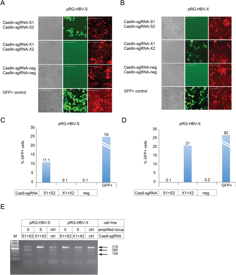
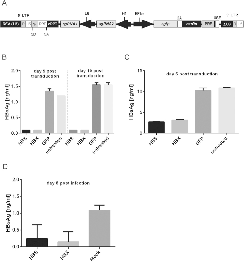
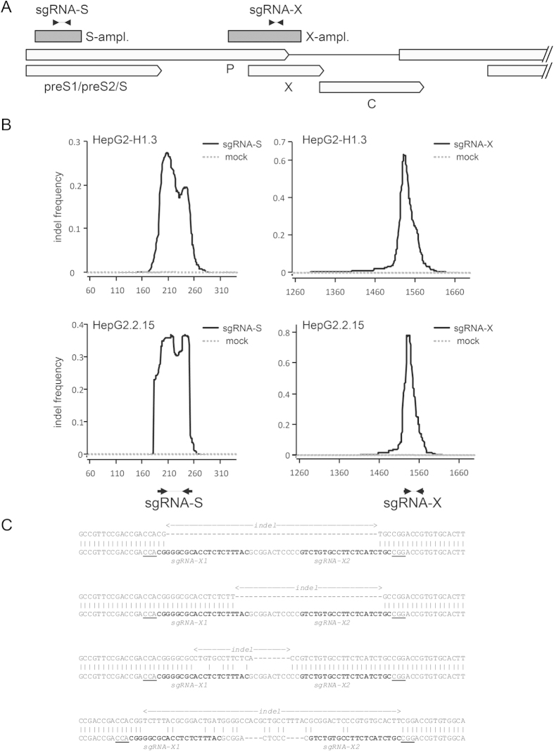
Current antiviral therapies cannot cure hepatitis B virus (HBV) infection; successful HBV eradication would require inactivation of the viral genome, which primarily persists in host cells as episomal covalently closed circular DNA (cccDNA) and, to a lesser extent, as chromosomally integrated sequences. However, novel designer enzymes, such as the CRISPR/Cas9 RNA-guided nuclease system, provide technologies for developing advanced therapy strategies that could directly attack the HBV genome. For therapeutic application in humans, such designer nucleases should recognize various HBV genotypes and cause minimal off-target effects. Here, we identified cross-genotype conserved HBV sequences in the S and X region of the HBV genome that were targeted for specific and effective cleavage by a Cas9 nickase. This approach disrupted not only episomal cccDNA and chromosomally integrated HBV target sites in reporter cell lines, but also HBV replication in chronically and de novo infected hepatoma cell lines. Our data demonstrate the feasibility of using the CRISPR/Cas9 nickase system for novel therapy strategies aiming to cure HBV infection.


Conflict of interest statement

M.K., F.B., J.S.z.W. and J.H. have filed a patent application based on the findings reported in this manuscript.

Figures

Figure 1
Figure 1. HBV-specific gRNA target sites for Cas9n recruitment.
(A) Schematic representation of the hepatitis B virus genome. Relaxed circular DNA (rcDNA) of the HB virion (thin continuous line), which is converted to cccDNA (thin continuous and dotted line) following hepatocyte infection, is indicated in the centre of the map. The four viral transcripts of the core (C), polymerase (P), and surface (S) and X proteins are indicated around the outside. Regions targeted by Cas9n via guide RNA (gRNA) specific to S and X sequences are indicated by arrows and scissors (scissors were drawn by Niklas Beschorner). (B) DNA sequence and sequence conservation of the regions targeted by Cas9n within the S and X gene of HBV. The sequence shown is based on genotype A consensus. Target sequences in ORF S and X are depicted (S1 and S2, or X1 and X2), each encompassing proto-spacer adjacent motifs (PAM, bold and boxed), 2 × 20 nucleotides complementary to gRNA (boxed) and offset distance between the two sequences complementary to gRNA (underlined).
Figure 2
Figure 2. Target site validation by transient transfection of HeLa cells.
(A) The HBV reporter plasmid for detecting Cas9n nuclease activity on HBV S or X target sequences is depicted at the top. The reporter construct contains a constitutive CMV promoter and sequences encoding RFP (red fluorescent protein) and GFP (enhanced green fluorescent protein), the latter lacking a start codon and positioned out of frame. RFP and GFP sequences are separated by the HBV S or X target site. Each target sequence contains two 20 bp regions necessary for gRNA binding and PAM motifs (expanded region). Cas9n nuclease activity aided by the pair of sgRNAs leads to two individual single stranded breaks within the target sequence (indicated by the large arrow; Cas9n cleaves the target sequence 3 nt upstream of the PAM region) which, upon non-homologous end joining repair, can lead to subtle sequence deletions and thereby frame shifts of the downstream GFP-specific sequence. (B) Cas9n activity on HBV sequences in HeLa cells at 48 h post transfection. Cultures were transfected with HBV S- or X-specific reporter and two Cas9n/sgRNA expression plasmids; HBV reporter plasmids alone (control); or with vectors expressing RFP, GFP and BFP (transfection control). Arrows indicate GFP expressing cells due to Cas9n activity. Scale bar = 400 μm. (C) Percentage of the total GFP + population in cultures containing the indicated vectors. Transfection control measured the presence of the positive control plasmid (GFP  +) by flow cytometry.
Figure 3
Figure 3. Analysis of Cas9n activity in HEK293 cells.
(A) HEK293 cells were transfected with the respective HBV reporter (pRG-HBV-S or pRG-HBV-X) and two plasmids for Cas9n/sgRNA expression (Cas9n-sgRNA-S1 and Cas9n-sgRNA-S2; Cas9n-sgRNA-X1 and Cas9n-sgRNA-X2). For transfection control, cells were transfected with a constitutively GFP-expressing plasmid or mock-transfected. Cells were imaged at 24 h post transfection. Arrows indicate GFP expressing cells due to Cas9n activity. Scale bar = 400 μm. (B) Cells were analyzed by FACS for RFP fluorescence to determine the presence of the HBV reporter in the transfected cells. (C,D) Target-specific Cas9n/gRNA nuclease activity was quantified by FACS analysis of GFP + cells within the populations of RFP + (reporter containing) cells.
Figure 4
Figure 4. Analysis of Cas9n activity by T7 endonuclease I assay.
(A) T7EI assays were performed using PCR primers (indicated by arrows) flanking the HBV S or X sequence in the respective reporter plasmid. (B) Detection of Cas9n-specific activity was visualized by gel electrophoresis. HEK293 cells were transfected as before and total genomic DNA was isolated at 72 h post transfection for subsequent T7EI cleavage. Arrows depict the sizes of wild-type and Cas9n-mutagenized DNA fragments. (C) T7EI assay using genomic DNA from GFP + HEK293 cell cultures at 24 h post transfection. (D) Sequence analysis of corresponding DNA samples. Alignment to the wild-type ORF S reporter sequence is shown. gRNA sequences (boxed), PAM (boxed and bold) and Cas9n-mediated deletions are indicated.
Figure 5
Figure 5. Targeting chromosomally integrated HBV-sequences using the CRISPR/Cas9n system in HEK293 cell cultures.
(A) Fluorescence microscopy images of the stable HBV S-specific reporter cell line cotransfected with plasmids expressing Cas9n and S or X (control) sequence-specific sgRNAs, or sgRNA targeted to an unrelated locus (negative control; Cas9n-sgRNA-neg). A constitutively GFP-expressing vector served as a transfection control. GFP expressing cells indicate Cas9n activity. Scale bar = 400 μm. (B) Analysis of a stable HBV X-specific reporter cell line as above. (C) HBV S sequence-specific Cas9n/sgRNA-mediated nuclease activity was quantified by GFP-specific FACS analysis. (D) As in C to quantify HBV X sequence-specific Cas9n/sgRNA-mediated nuclease activity.
Figure 6
Figure 6. Analysis of integrated HBV reporter constructs in HeLa cells.
(A) Targeting of chromosomally integrated HBV ORF S sequences by the CRISPR/Cas9n system was analyzed at 72 h post transfection, as described in Fig. 5A. GFP expressing cells indicate Cas9n activity. Scale bar = 400 μm. (B) Analysis of chromosomally integrated HBV ORF X sequences as described in Fig. 5B. (C,D) Quantification of the experiments shown in panel A and B, respectively, by FACS analysis of GFP + cells. (E) The stable HBV S and X target site-specific HeLa cultures, transfected with the indicated combinations of Cas9n and sgRNA-expressing vectors, analyzed by the T7EI cleavage assay. Targeting of an arbitrary genomic locus by sgRNA specific to this region was used as a positive control (ctrl).
Figure 7
Figure 7. Inactivation of HBV in chronically and de novo infected hepatocytes.
(A) The backbone of the HIV-derived lentiviral vector (LV) for delivering Cas9n and a pair of sgRNAs contains self-inactivating (SIN) long terminal repeats (LTR:ΔU3, R, U5), a Rev response element (RRE), a central polypurine tract (cPPT), a woodchuck hepatitis virus post-regulatory element (PRE), SV40 polyadenylation enhancer elements (USE), splice donor (SD), splice acceptor (SA) and packaging signal (Ψ) sites. Expression of an eGFP-2A peptide-Cas9n fusion protein is regulated by the internal human elongation factor 1α (EF1α) promoter. Transcription of two sgRNAs, sgRNA1 or sgRNA2, is regulated by a U6 or H1 pol III promoter. (B,C) HBsAg in the filtered supernatants of HepG2.2.15 and HepG2-H1.3 cells was quantified by ELISA at day 5 and/or at day 10 post transduction with LV-HBS, LV-HBX or LV-GFP. Experiments were performed in duplicate. The lower limit of detection (Background + 3 x S.D.) was 0.9 ng/ml for HBsAg. All values are given as mean concentration ng/ml ± S.D. (D) HBsAG in the filtered supernatant of LV-HBS, LV-HBX or mock transduced HepG2hNTCP cells was quantified as described before at day 8 post HBV infection. Experiments were performed in quadruplicate.
Figure 8
Figure 8. Analysis of indels in sgRNA-treated HepG2-H1.3 and HepG2-H2.2.15 cells.
(A) Scheme of the HBV genome depicting the location of amplicons spanning the S- and X-specific sgRNA target regions. The two amplicons are shown as dark gray boxes labelled S-ampl. and X-ampl. Arrows shown at the top indicate the genomic location of gRNA target sequences. (B) Indel frequency as detected in S-amplicons (left) or X-amplicons (right) from HepG2-H1.3 (top panels) or HepG2.2.15 (bottom panels) cells. The graphs represent the frequency with which each individual amplicon’s nucleotide is affected by indels in sgRNA-expressing (solid black line) or mock transduced cells (gray dashed line). Nucleotide positions given on the x-axis indicate coordinates on the full-length HBV genome (accession JN664938). The locations of gRNA target sequences are indicated by arrows underneath the bottom panels. (C) Example alignments of indel amplicon reads from Cas9n/sgRNA-X-expressing HepG2-H1.3 (top sequence in each alignment) aligned to wild-type HBV amplicon sequences (bottom sequence in each alignment). Indel sites are indicated above each read. The sgRNA target sequences and PAM motifs appear in bold or underlined, respectively.

Similar articles

Cited by

References

    1. Liaw Y. F. & Chu C. M. Hepatitis B virus infection. Lancet 373, 582–592 (2009). - PubMed
    1. Gerlich W. H. Medical virology of hepatitis B: how it began and where we are now. Virol. J. 10, 239 (2013). - PMC - PubMed
    1. You C. R., Lee S. W., Jang J. W. & Yoon S. K. Update on hepatitis B virus infection. World J. Gastroenterol. 20, 13293–13305 (2014). - PMC - PubMed
    1. Ganem D. & Prince A. M. Hepatitis B virus infection—natural history and clinical consequences. N. Engl. J. Med. 350, 1118–1129 (2004). - PubMed
    1. Block T. M., Guo H., & Guo J. T. Molecular virology of hepatitis B virus for clinicians. Clin. Liver Dis. 11, 685–706, vii (2007). - PMC - PubMed

Publication types

MeSH terms

Substances