Comparative Study
. 2015 Sep 3;11(9):e1005132.
doi: 10.1371/journal.ppat.1005132. eCollection 2015 Sep.

Distinct but Spatially Overlapping Intestinal Niches for Vancomycin-Resistant Enterococcus faecium and Carbapenem-Resistant Klebsiella pneumoniae

Affiliations
Comparative Study

Distinct but Spatially Overlapping Intestinal Niches for Vancomycin-Resistant Enterococcus faecium and Carbapenem-Resistant Klebsiella pneumoniae

Silvia Caballero et al. PLoS Pathog. .

Abstract

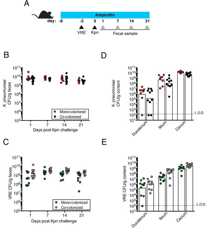
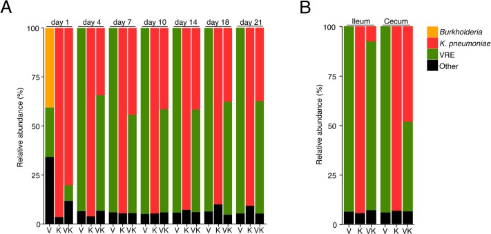
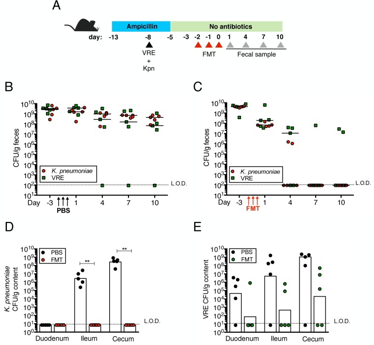
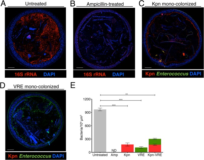
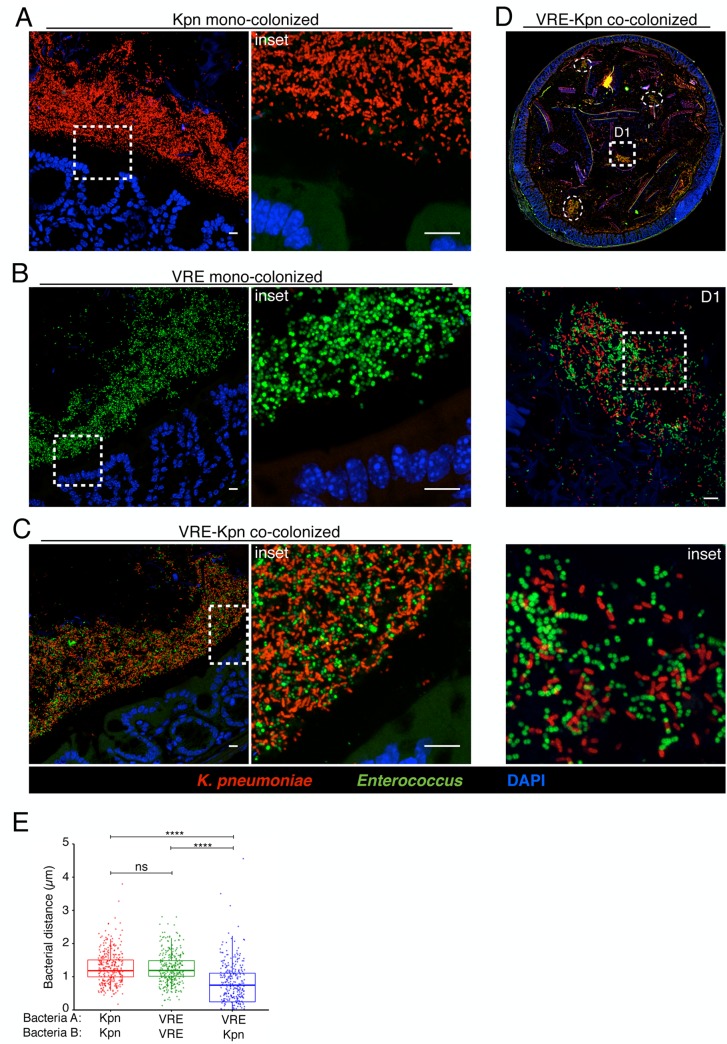
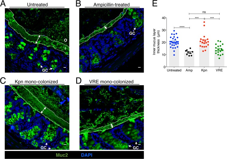
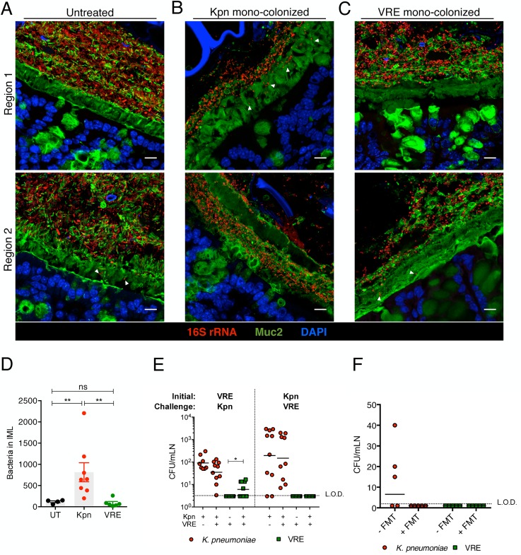
Antibiotic resistance among enterococci and γ-proteobacteria is an increasing problem in healthcare settings. Dense colonization of the gut by antibiotic-resistant bacteria facilitates their spread between patients and also leads to bloodstream and other systemic infections. Antibiotic-mediated destruction of the intestinal microbiota and consequent loss of colonization resistance are critical factors leading to persistence and spread of antibiotic-resistant bacteria. The mechanisms underlying microbiota-mediated colonization resistance remain incompletely defined and are likely distinct for different antibiotic-resistant bacterial species. It is unclear whether enterococci or γ-proteobacteria, upon expanding to high density in the gut, confer colonization resistance against competing bacterial species. Herein, we demonstrate that dense intestinal colonization with vancomycin-resistant Enterococcus faecium (VRE) does not reduce in vivo growth of carbapenem-resistant Klebsiella pneumoniae. Reciprocally, K. pneumoniae does not impair intestinal colonization by VRE. In contrast, transplantation of a diverse fecal microbiota eliminates both VRE and K. pneumoniae from the gut. Fluorescence in situ hybridization demonstrates that VRE and K. pneumoniae localize to the same regions in the colon but differ with respect to stimulation and invasion of the colonic mucus layer. While VRE and K. pneumoniae occupy the same three-dimensional space within the gut lumen, their independent growth and persistence in the gut suggests that they reside in distinct niches that satisfy their specific in vivo metabolic needs.


Conflict of interest statement

The authors have declared that no competing interests exist.

Figures

Fig 1
Fig 1. Pre-colonization with VRE does not prevent colonization by K. pneumoniae.
(A) Experimental design. Mice were treated with ampicillin for 29 days. On day 5 of ampicillin treatment, mice were inoculated with 5x10⁴ colony-forming units (CFU) of VRE by oral gavage or left uninfected. Three days later, half of the VRE-infected mice and the uninfected group were challenged with 5x10⁴ CFU of K. pneumoniae (Kpn). (B, C) CFU of K. pneumoniae (B) and VRE (C) were quantified in fecal pellets collected at different time points post K. pneumoniae inoculation. (D, E) Mice were sacrificed 21 days post K. pneumoniae challenge. K. pneumoniae (D) and VRE (E) burden was quantified in the luminal contents from the duodenum, ileum and cecum. L.O.D., limit of detection. Data were pooled from two independent experiments (n = 10 per group). (B-E) Data were analyzed by the Mann-Whitney test.
Fig 2
Fig 2. Pre-colonization with K. pneumoniae does not prevent colonization by VRE.
(A) Experimental design. Mice were treated with ampicillin for 29 days. On day 5 of ampicillin treatment, mice were inoculated with 5x10⁴ colony-forming units (CFU) of K. pneumoniae (Kpn) by oral gavage o left uninfected. Three days later, half of the K. pneumoniae-infected mice and the uninfected group were challenged with 5x10⁴ CFU of VRE. (B, C) CFU of VRE (B) and K. pneumoniae (C) were quantified in fecal pellets collected at different time points post VRE inoculation. (D, E) Mice were sacrificed 21 days post VRE challenge. VRE (D) and K. pneumoniae (E) burden was quantified in the luminal contents from the duodenum, ileum and cecum. L.O.D., limit of detection. Data were pooled from two independent experiments (n = 10 per group). (B-E) ****P<0.0001, by the the Mann-Whitney test.
Fig 3
Fig 3. K. pneumoniae and VRE achieve similar densities in the large intestine of co-colonized mice.
Ampicillin-treated mice were inoculated with K. pneumoniae by oral gavage or left uninfected. Three days later, half of the K. pneumoniae-infected mice and the uninfected group were challenged with VRE. Microbiota composition of mice colonized with VRE alone (V), K. pneumoniae alone (K) or both (VK) was determined by sequencing of the V4-V5 region of the 16S rRNA genes. (A) Fecal microbiota composition at different time points post VRE challenge. (B) Ileal and cecal microbiota composition at day 21 of colonization. (A,B) Each stacked bar represents the average of five individually-housed mice per time point.
Fig 4
Fig 4. Fecal bacteriotherapy eliminates established K. pneumoniae and VRE intestinal domination.
(A) Experimental design. Mice were treated with ampicillin for 8 days. On day 5 of ampicillin treatment, mice were simultaneously infected with 5x10⁴ CFU of VRE and K. pneumoniae (Kpn). Three days post infection, ampicillin treatment was stopped. Mice were administered PBS or a fecal microbiota transplant (FMT) from an untreated mouse on three consecutive days starting on the third day after ampicillin cessation. (B, C) VRE and K. pneumoniae burden was quantified in fecal pellets at the indicated time points after the last PBS (B) or FMT (C) dose. (D, E) PBS- and FMT-treated mice were sacrificed on day 10 following the last treatment dose and numbers of K. pneumoniae (D) and VRE (E) CFU were quantified in the duodenum, ileum and cecum. L.O.D., limit of detection. n ≥ 5 per group. (B-E) **P<0.005 by the Mann-Whitney test.
Fig 5
Fig 5. K. pneumoniae and VRE occupy a fraction of the total available space in the colon.
(A-E) Visualization of bacterial localization by FISH. Entire colon cross-sections from untreated mice (A) and mice treated with ampicillin for 3 weeks (B) were stained with a universal probe that targets the 16S rRNA gene of all bacteria. Cross-sections from ampicillin-treated mice colonized with K. pneumoniae (C) or VRE (D) for 21 days were hybridized with probes specific for K. pneumoniae (Kpn) and Enterococcus, respectively. Sections were counterstained with Hoechst dye to visualize nuclei. Images are representative of 5 mice per group. Scale bar, 500 μm. (E) Number of bacteria per unit area of whole colon cross-sections. n = 3 per group. ND = non-detectable. Error bars (mean ± SEM). **P<0.005, ***P<0.0005 by the Mann-Whitney test.
Fig 6
Fig 6. K. pneumoniae and VRE reside within the same intestinal regions but occupy distinct metabolic niches.
(A-D) Spatial localization of K. pneumoniae and VRE in the colon. Colon sections from ampicillin-treated mice colonized for 21 days with K. pneumoniae alone (A), VRE alone (B) and K. pneumoniae together with VRE (C, D) were hybridized with probes specific for K. pneumoniae and Enterococcus. (D) VRE and K. pneumoniae islands (dashed circles and square) in the colonic lumen of co-colonized mice. (A-D) All sections were counterstained with Hoechst dye to visualize nuclei. Scale bars, 10 μm. Insets, 63X oil objective plus 4X digital zoom. Images are representative of at least 5 mice per group. (E) Minimum distance between neighboring bacteria determined by confocal microscopy. ns = non-significant; ****P<0.0001, by the Mann-Whitney test.
Fig 7
Fig 7. K. pneumoniae and VRE colonization influences the thickness of the inner mucus layer.
(A-D) Colon sections from untreated mice (A), ampicillin-treated mice (B) and ampicillin-treated mice mono-colonized with either K. pneumoniae (C) or VRE (D) for 21 days were stained with an anti-Muc2 antibody to visualize the inner and outer mucus layers along with goblet cells (arrows). Double arrows denote the inner mucus layer. i, inner mucus layer; o, outer mucus layer; GC, goblet cell. Scale bars, 10 μm. Images are representative of 5 mice per group. (E) Quantification of inner mucus layer thickness. Error bars (mean ± SEM). n = 3–6 mice per group. ns = non-significant; ***P<0.0005, ****P<0.0001, by the unpaired Student t test.
Fig 8
Fig 8. Differential mucus layer infiltration and translocation by VRE and K. pneumoniae.
(A-C) Dual immunostaining of colon sections from untreated mice (A) and ampicillin-treated mice mono-colonized with either K. pneumoniae (B) or VRE (C) for 21 days using anti-Muc2 and a pan-bacterial 16S rRNA gene FISH probe. Sections were counterstained with Hoechst dye to visualize nuclei. Arrowheads indicate bacteria within the inner mucus layer. Scale bar, 10 μm. Images are representative of 5 mice per group. Boundaries of the inner mucus layer (IML) zone were determined by the density of Muc2 staining and the stratified organization characteristic of the inner, but not outer, mucus layer. (D) Number of bacteria within the IML. UT, untreated. (E) Numbers of VRE and K. pneumoniae in mesenteric lymph nodes of mono-colonized mice and mice pre-colonized with either VRE or K. pneumoniae (initial strain) and challenged with the opposite strain at day 21 post challenge. Data were pooled from two independent experiments. (F) Numbers of VRE and K. pneumoniae in mesenteric lymph nodes of mice co-colonized with VRE and K. pneumoniae with or without a fecal transplant (FMT) 10 days after receiving the last of three FMT/PBS doses. Data were pooled from two independent experiments. L.O.D., limit of detection. (D-F) ns = non-significant; *P<0.05, **P<0.005, by the Mann-Whitney test.

Similar articles

Cited by

References

    1. Magill SS, Edwards JR, Bamberg W, Beldavs ZG, Dumyati G, Kainer MA, et al. Multistate point-prevalence survey of health care-associated infections. N Engl J Med 2014, March 27;370(13):1198–208. 10.1056/NEJMoa1306801 - DOI - PMC - PubMed
    1. Arias CA, Murray BE. Emergence and management of drug-resistant enterococcal infections. Expert Rev Anti Infect Ther 2008, October;6(5):637–55. 10.1586/14787210.6.5.637 - DOI - PubMed
    1. Gupta N, Limbago BM, Patel JB, Kallen AJ. Carbapenem-resistant enterobacteriaceae: Epidemiology and prevention. Clin Infect Dis 2011, July 1;53(1):60–7. 10.1093/cid/cir202 - DOI - PubMed
    1. Safdar N, Maki DG. The commonality of risk factors for nosocomial colonization and infection with antimicrobial-resistant staphylococcus aureus, enterococcus, gram-negative bacilli, clostridium difficile, and candida. Ann Intern Med 2002, June 4;136(11):834–44. - PubMed
    1. Donskey CJ. The role of the intestinal tract as a reservoir and source for transmission of nosocomial pathogens. Clin Infect Dis 2004, July 15;39(2):219–26. - PubMed

Publication types

MeSH terms