Comparative Study
. 2015 Sep 3;10(9):e0137243.
doi: 10.1371/journal.pone.0137243. eCollection 2015.

Differential Subcellular Localization of Leishmania Alba-Domain Proteins throughout the Parasite Development

Affiliations
Comparative Study

Differential Subcellular Localization of Leishmania Alba-Domain Proteins throughout the Parasite Development

Aurélien Dupé et al. PLoS One. .

Abstract

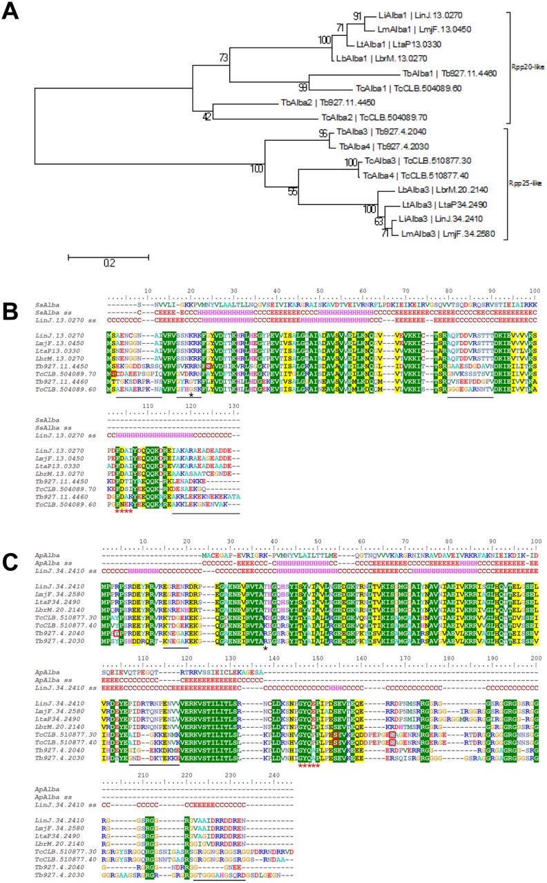
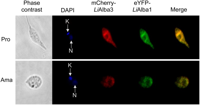
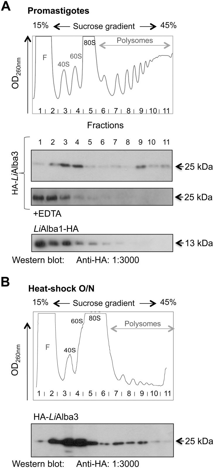
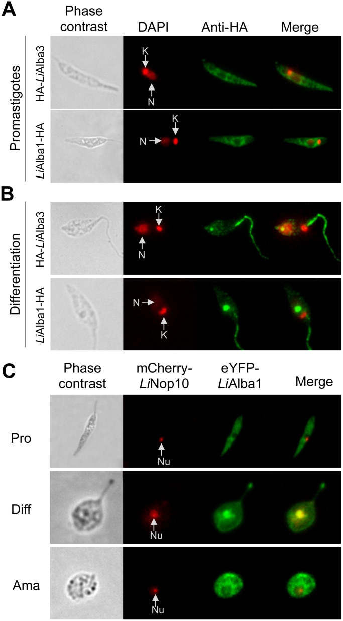
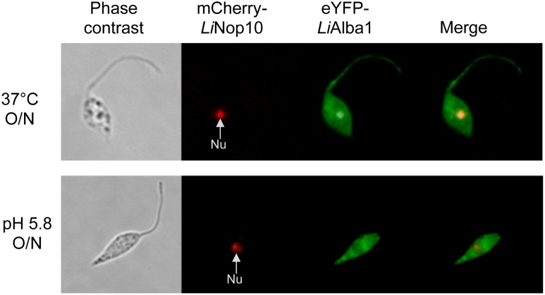
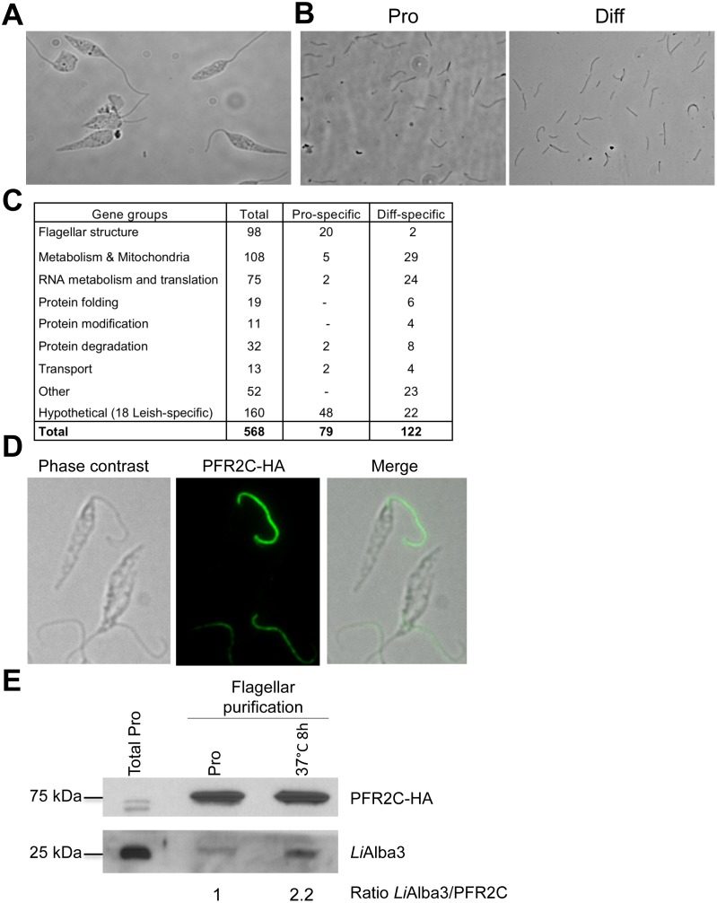
Alba-domain proteins are RNA-binding proteins found in archaea and eukaryotes and recently studied in protozoan parasites where they play a role in the regulation of virulence factors and stage-specific proteins. This work describes in silico structural characterization, cellular localization and biochemical analyses of Alba-domain proteins in Leishmania infantum. We show that in contrast to other protozoa, Leishmania have two Alba-domain proteins, LiAlba1 and LiAlba3, representative of the Rpp20- and the Rpp25-like eukaryotic subfamilies, respectively, which share several sequence and structural similarities but also important differences with orthologs in other protozoa, especially in sequences targeted for post-translational modifications. LiAlba1 and LiAlba3 proteins form a complex interacting with other RNA-binding proteins, ribosomal subunits, and translation factors as supported by co-immunoprecipitation and sucrose gradient sedimentation analysis. A higher co-sedimentation of Alba proteins with ribosomal subunits was seen upon conditions of decreased translation, suggesting a role of these proteins in translational repression. The Leishmania Alba-domain proteins display differential cellular localization throughout the parasite development. In the insect promastigote stage, Alba proteins co-localize predominantly to the cytoplasm but they translocate to the nucleolus and the flagellum upon amastigote differentiation in the mammalian host and are found back to the cytoplasm once amastigote differentiation is completed. Heat-shock, a major signal of amastigote differentiation, triggers Alba translocation to the nucleolus and the flagellum. Purification of the Leishmania flagellum confirmed LiAlba3 enrichment in this organelle during amastigote differentiation. Moreover, partial characterization of the Leishmania flagellum proteome of promastigotes and differentiating amastigotes revealed the presence of other RNA-binding proteins, as well as differences in the flagellum composition between these two parasite lifestages. Shuttling of Alba-domain proteins between the cytoplasm and the nucleolus or the flagellum throughout the parasite life cycle suggests that these RNA-binding proteins participate in several distinct regulatory pathways controlling developmental gene expression in Leishmania.


Conflict of interest statement

Competing Interests: The authors have declared that no competing interests exist.

Figures

Fig 1
Fig 1. Sequence alignment and phylogeny of Alba-domain proteins in Leishmania, Trypanosoma brucei and T. cruzi species.
(A) Neighbor-joining tree showing the phylogenetic relationship between the Alba-domain proteins of TriTryps. Evolutional distances (scale) were estimated as the number of amino acid substitutions per site, considering Poisson correction. The two subgroups Rpp20-like and Rpp25-like are marked. (B) ClustalW alignment of Rpp20-like Alba-domain proteins merged with the in silico structure prediction of LiAlba1 (LinJ.13.0270) using the Phyre algorithm. The best score was obtained with the Alba protein from Sulfolobus solfataricus (Ss)(NCBI WP_010923153.1). ss: secondary structure in silico prediction; C: coil; H: helix; E: Sheet. Red squares indicate amino acids known to be phosphorylated on these specific genes (T. cruzi and T. brucei). The black star shows the expected position for Sir2 acetylation and the red stars underline the signature motif of the subgroup. Sequence variations in coiled regions between Trypanosoma spp. and Leishmania spp. are underlined with a black bar. LiAlba1 was used for Phyre structure prediction. (C) As in B for the Rpp25-like Alba-domain proteins. Structure prediction of LiAlba3 using Phyre outputs Alba2 from Aeropyrum pernix (Ap) K1 (NCBI WP_010866616.1) as the best match. LiAlba3 (LinJ.34.2410) was used for Phyre structure prediction. LinJ: L. infantum; LmjF: L. major; LtaP: L. tarentolae; LbrM: L. braziliensis; Tb: T. brucei; Tc: T. cruzi.
Fig 2
Fig 2. Alba-domain proteins co-localize to the cytoplasm of promastigote and amastigote Leishmania life stages.
Direct fluorescence images of recombinant L. infantum promastigotes (Pro) and axenic amastigotes (Ama) at passage 4 co-expressing eYPF-LiAlba1 (green) and mCh-LiAlba3 (red) proteins. Green and red pixels overlapped in the digital images yielding yellow/orange signals. The nucleus (N) and kinetoplastid DNA (K) were stained with DAPI (blue).
Fig 3
Fig 3. Alba-domain proteins are associated with ribosomal subunits.
Polysome fractionation of L. infantum expressing HA-tagged Alba1 and Alba3 proteins by 15–45% sucrose gradient was carried out using logarithmic phase promastigotes (26°C) (A) or heat-stressed parasites grown O/N at 37°C (B). Graphical representations present the RNA content of each collected fraction after ultracentrifugation on 15–45% sucrose gradient. F: Free RNA; 40S, 60S and 80S: ribosomal subunits and monosomes, respectively. Each fraction was loaded on 12% SDS-PAGE and transferred on a nylon membrane for Western blot analysis to detect HA-LiAlba3 and LiAlba1-HA proteins using an anti-HA antibody. As a control, half of the protein extracts were incubated with EDTA before ultracentrifugation to disrupt association of the polyribosomes with mRNAs.
Fig 4
Fig 4. Alba-domain proteins translocate from the cytoplasm to the flagellum and the nucleolus upon Leishmania amastigote differentiation.
Subcellular localization of LiAlba1-HA and HA-LiAlba3 proteins in promastigotes (A) and upon amastigote differentiation (8 h in MAA medium pH 5.8 at 37°C) (B) was assessed by indirect immunofluorescence studies using an anti-HA antibody as described in Materials and Methods. DAPI staining (red) allows detection of the nucleus (N) and kinetoplastid DNA (K). C) Immunofluorescence images of wild type L. infantum episomally co-expressing pSP-NEOalphaIR-eYPF-LiAlba1 and pSP-HYGalphaIR-mCh-LiNOP10 grown as promastigotes (Pro), differentiating amastigotes (Diff) and amastigotes (Ama). LiNOP10 was used as a nucleolar (Nu) control.
Fig 5
Fig 5. Heat stress triggers differential localization of Alba-domain proteins in Leishmania.
Immunofluorescence images for eYPF-LiAlba1 (green) and mCh-LiNOP10 (red) in L. infantum promastigotes co-expressing pSP-NEOalphaIR-eYPF-LiAlba1 and pSP-HYGalphaIR-mCh-LiNOP10 submitted to heat stress (from 25°C to 37°C) or to acidic pH (pH 5.8) O/N. Nu: nucleolus.
Fig 6
Fig 6. Flagellum purification demonstrates an enrichment of LiAlba3 in the Leishmania flagellum during heat stress.
Phase contrast images of intact L. infantum promastigotes (A) and of purified flagella after sucrose gradient isolation (B) as described in Materials and Methods. (C) Summary of MS/MS identified proteins from four independent experiments of flagellum purification (two from promastigote cell extracts and two from 8 h-differentiating amastigotes). Identified genes were classified according to their gene ontology and to characterized orthologs in Trypanosoma spp. based on GeneDB and TriTrypDB gene annotations. Only genes identified at least twice with a minimum of 2 peptides are shown here (see Tables 2, S3 and S4 for the complete list of the identified L. infantum flagellum proteins). (D) Confirmation of flagellar localization of PFR2C-HA in recombinant L. infantum expressing pSP-alphaIRNEOalphaIR-PFR2C-HA. (E) Western blot analysis and quantification of endogenous LiAlba3 in purified flagellum fractions upon promastigote conditions of growth (Pro) or following 8 h of temperature stress (37°C). After flagellum purification, flagella from promastigotes and heat-stressed parasites were counted on a Malassey (hemocytometer) to load an equivalent number of flagella on the gel. Total proteins of promastigote cells were loaded as a control. Relative quantification was performed using ImageJ blot.

References

    1. Herwaldt BL. Leishmaniasis. Lancet. 1999;354(9185):1191–9. - PubMed
    1. Bates PA. Transmission of Leishmania metacyclic promastigotes by phlebotomine sand flies. Int J Parasitol. 2007;37(10):1097–106. - PMC - PubMed
    1. Kaye P, Scott P. Leishmaniasis: complexity at the host-pathogen interface. Nat Rev Microbiol. 2011;9(8):604–15. 10.1038/nrmicro2608 - DOI - PubMed
    1. Zilberstein D, Shapira M. The role of pH and temperature in the development of Leishmania parasites. Annu Rev Microbiol. 1994;48:449–70. - PubMed
    1. Rochette A, Raymond F, Corbeil J, Ouellette M, Papadopoulou B. Whole-genome comparative RNA expression profiling of axenic and intracellular amastigote forms of Leishmania infantum . Mol Biochem Parasitol. 2009;165(1):32–47. 10.1016/j.molbiopara.2008.12.012 - DOI - PubMed

Publication types

LinkOut - more resources