

Retinal and Choroidal Folds in Papilledema
- PMID: 26335066
- PMCID: PMC4562343
- DOI: 10.1167/iovs.15-17459
Retinal and Choroidal Folds in Papilledema
Abstract
Purpose: To determine the frequency, patterns, associations, and biomechanical implications of retinal and choroidal folds in papilledema due to idiopathic intracranial hypertension (IIH).
Methods: Retinal and choroidal folds were studied in patients enrolled in the IIH Treatment Trial using fundus photography (n = 165 study eyes) and spectral-domain optical coherence tomography (SD-OCT; n = 125). We examined the association between folds and peripapillary shape, retinal nerve fiber layer (RNFL) thickness, disc volume, Frisén grade, acuity, perimetric mean deviation, intraocular pressure, intracranial pressure, and refractive error.
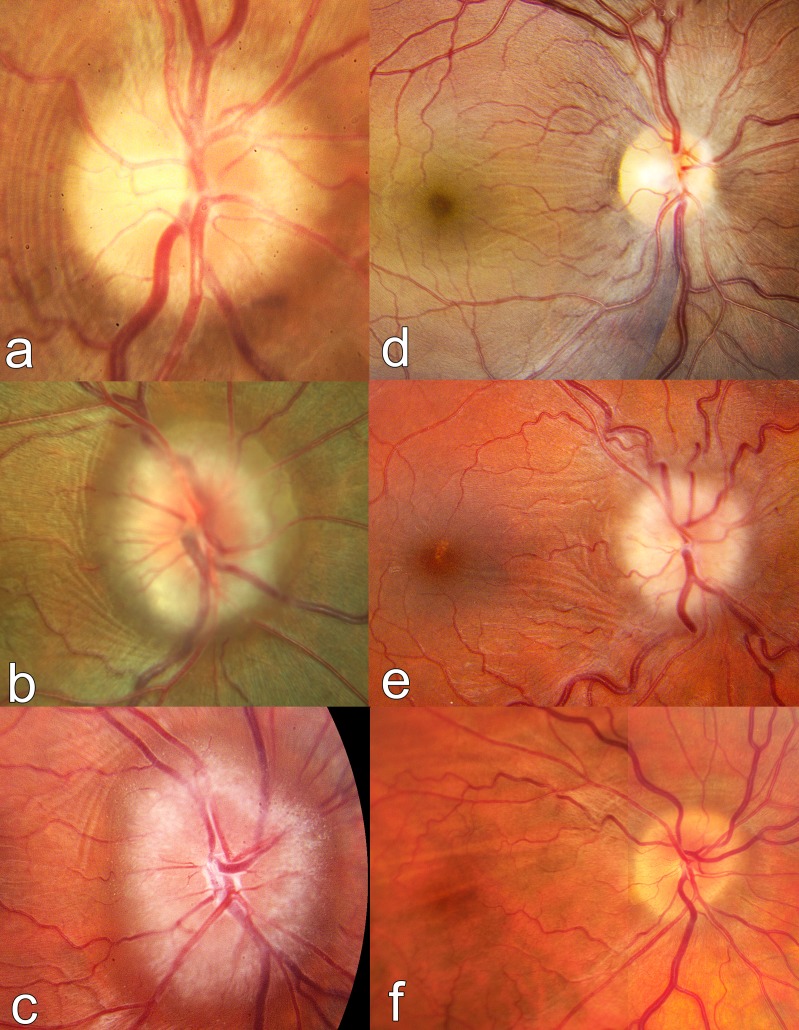
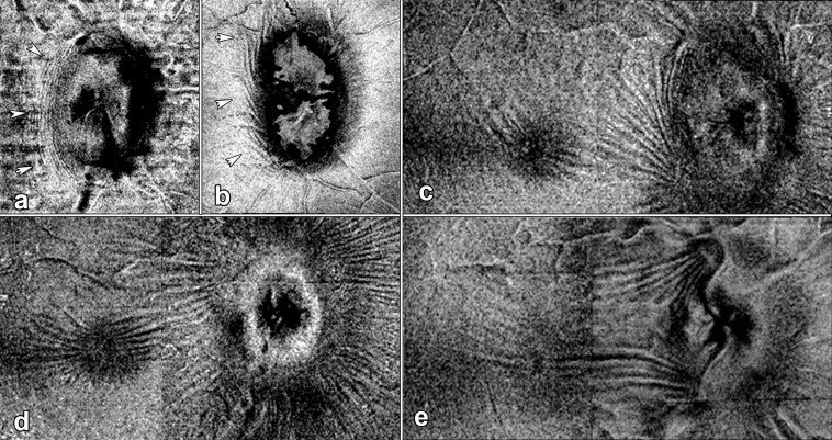
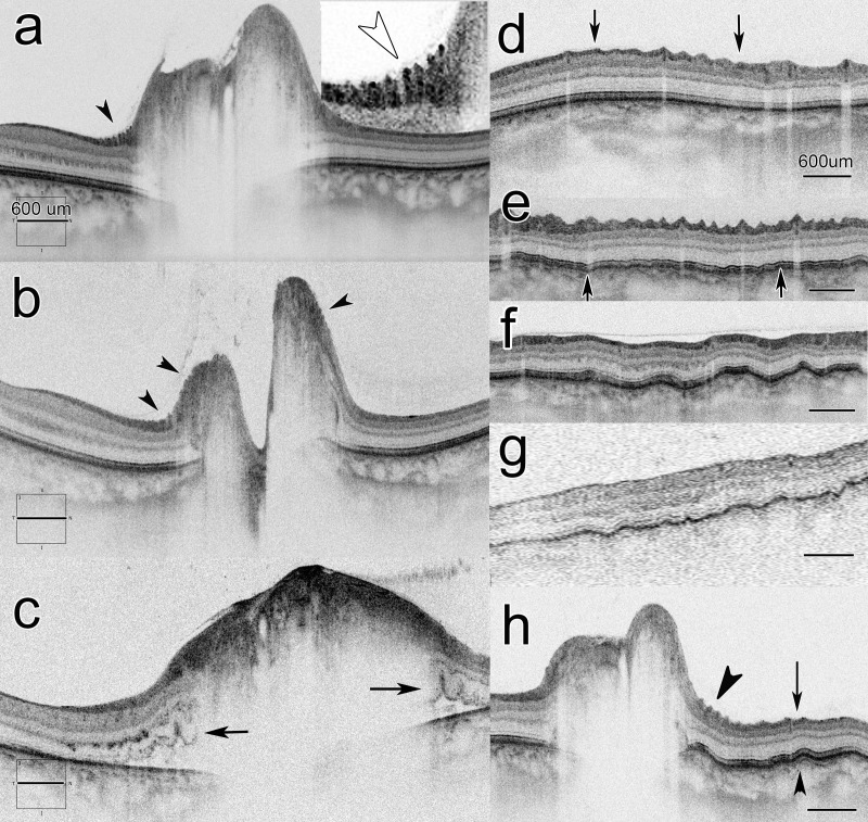
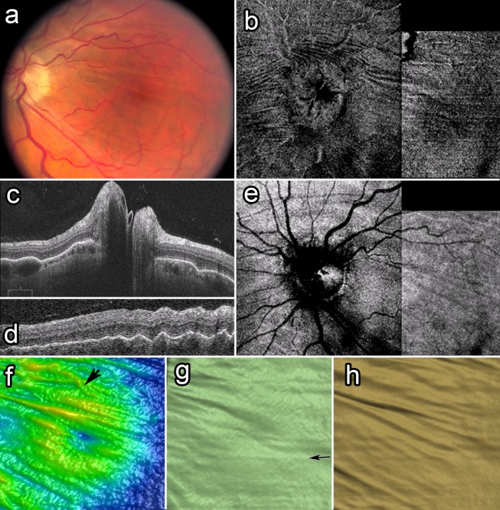
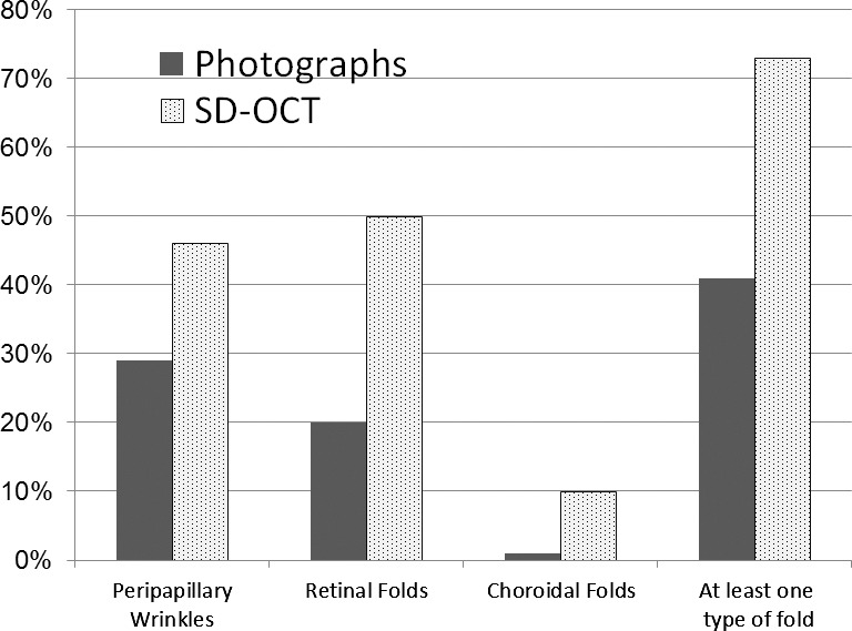
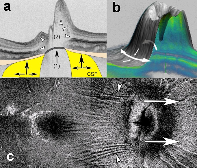
Results: We identified three types of folds in IIH patients with papilledema: peripapillary wrinkles (PPW), retinal folds (RF), and choroidal folds (CF). Frequency, with photos, was 26%, 19%, and 1%, respectively; SD-OCT frequency was 46%, 47%, and 10%. At least one type of fold was present in 41% of patients with photos and 73% with SD-OCT. Spectral-domain OCT was more sensitive. Structural parameters related to the severity of papilledema were associated with PPW and RF, whereas anterior deformation of the peripapillary RPE/basement membrane layer was associated with CF and RF. Folds were not associated with vision loss at baseline.
Conclusions: Folds in papilledema are biomechanical signs of stress/strain on the optic nerve head and load-bearing structures induced by intracranial hypertension. Folds are best imaged with SD-OCT. The patterns of retinal and choroidal folds are the products of a complex interplay between the degree of papilledema and anterior deformation of the load-bearing structures (sclera and possibly the lamina cribrosa), both modulated by structural geometry and material properties of the optic nerve head. (ClinicalTrials.gov number, NCT01003639.).
Figures
Comment in
-
Choroidal Folds in Astronauts.Invest Ophthalmol Vis Sci. 2016 Feb;57(2):592. doi: 10.1167/iovs.15-18720. Invest Ophthalmol Vis Sci. 2016. PMID: 26886892 No abstract available.
-
Author Response: Choroidal Folds in Astronauts.Invest Ophthalmol Vis Sci. 2016 Feb;57(2):593. doi: 10.1167/iovs.15-18927. Invest Ophthalmol Vis Sci. 2016. PMID: 26886893 No abstract available.
References
-
- Genzer JG. J. Soft matter with hard skin: from skin wrinkles to templating and material characterization. Soft Matter. 2006; 2: 310– 323. - PubMed
-
- Chung JY,, Nolte AJ,, Stafford CM. Surface wrinkling: a versatile platform for measuring thin-film properties. Adv Mater. 2011; 23: 3493– 68. - PubMed
-
- Yang S,, Khare, K,, Lin PC. Harnessing surface wrinkle patterns in soft matter. Adv Funct Mater. 2010; 20: 2550– 2564.
-
- Li B,, Cao, YP,, Feng XQ,, Gao H. Mechanics of morphological instabilities and surface wrinkling in soft materials: a review. Soft Matter. 2012; 8: 5728– 5745.
-
- Cerda E,, Mahadevan L. Geometry and physics of wrinkling. Phys Rev Lett. 2003. ; 90: 074302. - PubMed
Publication types
MeSH terms
Associated data
Grants and funding
LinkOut - more resources
Full Text Sources
Other Literature Sources
Medical
