. 2015 Sep 3;9(9):e0004067.
doi: 10.1371/journal.pntd.0004067. eCollection 2015.

Schistosoma mansoni Infection in Ugandan Men Is Associated with Increased Abundance and Function of HIV Target Cells in Blood, but Not the Foreskin: A Cross-sectional Study

Affiliations

Schistosoma mansoni Infection in Ugandan Men Is Associated with Increased Abundance and Function of HIV Target Cells in Blood, but Not the Foreskin: A Cross-sectional Study

Jessica L Prodger et al. PLoS Negl Trop Dis. .

Abstract

Background: Schistosoma mansoni infection has been associated with an increased HIV prevalence in humans and SHIV incidence in primate models. We hypothesized that immune activation from this gastrointestinal mucosa infection would increase highly HIV-susceptible CD4 T cell subsets in the blood and the foreskin through common mucosal homing.

Methodology/principal findings: Foreskin tissue and blood were obtained from 34 HIV- and malaria-uninfected Ugandan men who volunteered for elective circumcision, 12 of whom were definitively positive for S. mansoni eggs in stool and 12 definitively negative for both S. mansoni eggs and worm antigen. Tissue and blood T cell subsets were characterized by flow cytometry and immunohistochemistry (IHC). Th17 and Th1 cells from both the blood and foreskin expressed higher levels of CCR5 and were more activated than other CD4 T cell subsets. S. mansoni-infected men had a higher frequency of systemic Th1 cells (22.9 vs. 16.5% of blood CD4 T cells, p<0.05), Th17 cells (2.3 vs. 1.5%, p<0.05), and Th22 cells (0.5 vs. 0.3%, p<0.01) than uninfected men. Additionally, Th17 cells in the blood of S. mansoni-infected men demonstrated enhanced function (28.1 vs. 16.3% producing multiple cytokines, p = 0.046). However, these immune alterations were not observed in foreskin tissue.

Conclusions/significance: S. mansoni infection was associated with an increased frequency of highly HIV-susceptible Th1, Th17 and Th22 cell subsets in the blood, but these T cell immune differences did not extend to the foreskin. S. mansoni induced changes in T cell immunology mediated through the common mucosal immune system are not likely to increase HIV susceptibility in the foreskin.


Conflict of interest statement

The authors have declared that no competing interests exist.

Figures

Fig 1
Fig 1. Flow cytometry gating strategy for Th subsets isolated from (A) the blood and (B) the foreskin.
Blood cells were isolated by density centrifugation, and foreskin cells by mechanical and enzymatic digestion of tissue. Dead cells and doublets were excluded. Lymphocytes were identified based on characteristic size and granularity in blood samples and gates applied to foreskin samples. CD4 T cells were identified by expression of CD3 and CD4. Th subsets among CD4 T cells were identified by cytokine production in response to non-specific stimuli (SEB shown). Gates for cytokine production were based on unstimulated samples. Th17 cells were identified by production of IL-17A; Th1 cells by production of IFNγ and not IL-17A; Th22 cells by production of IL-22 in the absence of either IFNγ or IL-17A. Representative plots showing expression of CCR5, CD69 and HLA-DR on CD4 T cells are also shown.
Fig 2
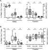
Fig 2. Differential expression of markers of HIV susceptibility on Th populations isolated from the blood and foreskin.
Expression of (A) CCR5, (B) CD69, and (C) HLA-DR was measured on CD4 T cells isolated from the blood (PBMCs) and foreskin tissue using flow cytometry. “Other” refers to CD4+/CD3+ cells that do not produce IL-17A, IFNγ or IL-22. Expression of surface markers on Th subsets was compared using the Friedman chi-square test; post-hoc pairwise comparisons made using Wilcoxon related samples rank test and Bonferroni adjusted p-values are reported (*p<0.05; **p<0.01; ***p<0.001).
Fig 3
Fig 3. HIV target cells in the blood and foreskin of men infected with S. mansoni.
HIV target cells were identified by flow cytometry from cells isolated from the blood (PBMC) or foreskin, and were compared between men shedding S. mansoni eggs (hatched bars) and men who were free of Schistosoma infection (clear bars). (A) Proportions of CD4 T cells that are Th1, Th17 or Th22 cells. (B) Expression of markers of HIV susceptibility on CD4 T cells. T cell populations were compared between S. mansoni-infected and uninfected men by Mann-Whitney U test (*p<0.05; **p<0.01).
Fig 4
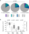
Fig 4. Functional capacity of Th17 cells isolated from the blood of men infected with S. mansoni.
The ability of Th17 cells isolated from the blood to produce a second or third cytokine (IL-22 and/or IFNγ in addition to IL-17A) in response to non-specific stimulation (PMA/iono) was measured using flow cytometry. (A) Proportion of Th17 cells producing additional cytokines was compared between men infected with S. mansoni (shedding eggs), intermediate (presence of worms detected by Urine-CCA, but not shedding eggs), and uninfected men (no detection of worm antigen or eggs). Assessment of polyfunctionality was performed using SPICE v5.35 software with polyfunctionality compared between S. mansoni-infected and uninfected men using a chi-squared distribution (1000 permutations of pies)[63]. (B) Post-hoc comparison of individual functional subsets between S. mansoni-infected (hatched bars) and uninfected men (related samples Wilcoxon rank test).

Similar articles

Cited by

References

    1. UNAIDS. Global Report: UNAIDS report on the global AIDS epidemic 2013. WHO Library Cataloguing-in-Publication Data: 2013.
    1. Keele BF, Giorgi EE, Salazar-Gonzalez JF, Decker JM, Pham KT, Salazar MG, et al. Identification and characterization of transmitted and early founder virus envelopes in primary HIV-1 infection. Proc Natl Acad Sci U S A. 2008;105(21):7552–7. Epub 2008/05/21. 10.1073/pnas.0802203105 - DOI - PMC - PubMed
    1. Hladik F, McElrath MJ. Setting the stage: host invasion by HIV. Nat Rev Immunol. 2008;8(6):447–57. Epub 2008/05/13. 10.1038/nri2302 - DOI - PMC - PubMed
    1. Yi TJ, Shannon B, Prodger J, McKinnon L, Kaul R. Genital immunology and HIV susceptibility in young women. Am J Reprod Immunol. 2013;69 Suppl 1:74–9. 10.1111/aji.12035 - DOI - PubMed
    1. Talbert-Slagle K, Atkins KE, Yan KK, Khurana E, Gerstein M, Bradley EH, et al. Cellular superspreaders: an epidemiological perspective on HIV infection inside the body. PLoS Pathog. 2014;10(5):e1004092 10.1371/journal.ppat.1004092 - DOI - PMC - PubMed

Publication types