Review
. 2015 Sep 2;87(5):946-61.
doi: 10.1016/j.neuron.2015.08.001.

Neural Control of Breathing and CO2 Homeostasis

Affiliations
Review

Neural Control of Breathing and CO2 Homeostasis

Patrice G Guyenet et al. Neuron. .

Abstract

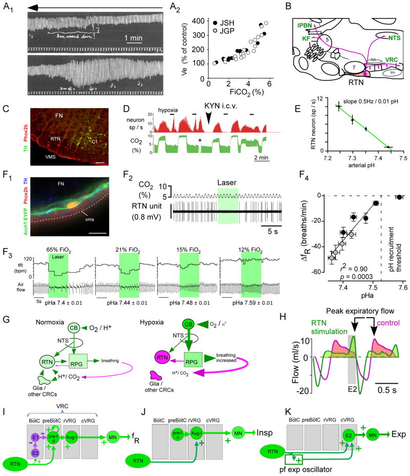
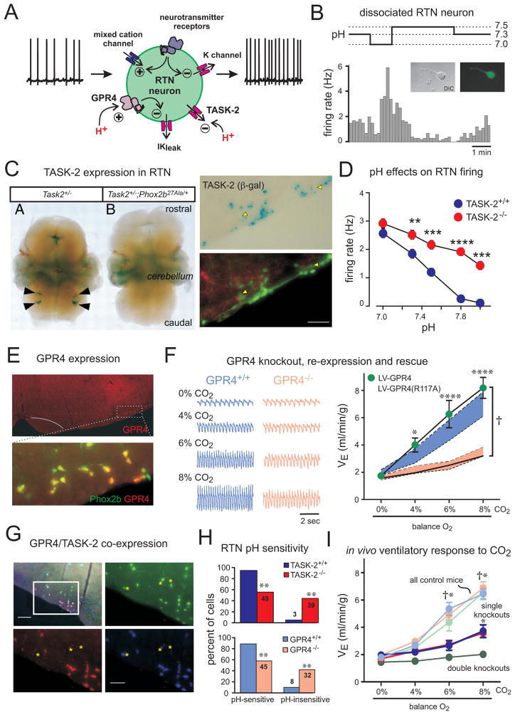
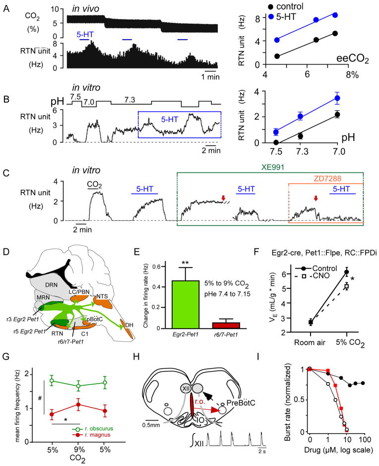
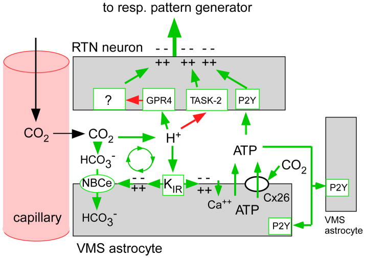
Recent advances have clarified how the brain detects CO2 to regulate breathing (central respiratory chemoreception). These mechanisms are reviewed and their significance is presented in the general context of CO2/pH homeostasis through breathing. At rest, respiratory chemoreflexes initiated at peripheral and central sites mediate rapid stabilization of arterial PCO2 and pH. Specific brainstem neurons (e.g., retrotrapezoid nucleus, RTN; serotonergic) are activated by PCO2 and stimulate breathing. RTN neurons detect CO2 via intrinsic proton receptors (TASK-2, GPR4), synaptic input from peripheral chemoreceptors and signals from astrocytes. Respiratory chemoreflexes are arousal state dependent whereas chemoreceptor stimulation produces arousal. When abnormal, these interactions lead to sleep-disordered breathing. During exercise, central command and reflexes from exercising muscles produce the breathing stimulation required to maintain arterial PCO2 and pH despite elevated metabolic activity. The neural circuits underlying central command and muscle afferent control of breathing remain elusive and represent a fertile area for future investigation.


Figures

Figure 1
Figure 1. the retrotrapezoid nucleus, RTN
(A1) the hypercapnic ventilatory reflex in humans (smoked drum recording to be read from right to left, top to bottom). Fraction inspired CO2 (FiCO2) was gradually increased by rebreathing air from box placed around head (reproduced from (Haldane and Priestley, 1905)). Amplitude of signal represents Vt (tidal volume). (A2) Plot of % increase in Ve (Vt x fR) vs. FiCO2 from Table on page 249 of (Haldane and Priestley, 1905)(JSH, Haldane; JGP, Priestly). (B) Location and projections of the rodent RTN (parasagittal section). Abbreviations: KF, Kölliker-Fuse nucleus; lPBN, lateral parabrachial nuc.; lrn, lateral reticular nuc.; NTS, solitary tract nuc.; VRC, ventral respiratory column; 5, trigeminal motor nuc.; 7, facial nuc.; (C) Transverse section at bregma level −11.5mm of an adult rat (left side) showing RTN neurons (FN: facial motor nucleus). The Phox2b+:tyrosine-hydroxylase (TH) neurons are RTN neurons. The Phox2b+:TH+ are C1 adrenergic neurons. Cal: 0.1 mm. From (Guyenet, 2008) (D) single RTN neuron recorded in an anesthetized rat. The firing frequency of the neuron (upper trace) is increased both by raising FiCO2 in a background of hyperoxia (selective central chemoreceptor activation), by brief hypoxia (selective carotid body stimulation) or by short asphyxia (asterisk); the top of the CO2 trace (CO2 concentration at end-expiration) approximates arterial PCO2 measured in % of atmospheric pressure. Blocking excitatory transmission with kynurenic acid i.c.v. eliminates the input from the carotid body but the effect of hypercapnia is unchanged (adapted from (Mulkey et al., 2004). (E) Relationship between discharge rate of RTN neurons (N=11) and arterial pH in anesthetized rats (glutamatergic transmission blocked with kynurenate as in D; adapted from (Guyenet et al., 2005)). (F1) Rat RTN neuron transduced with archaerhodopsin-eYFP (ArchT). Cal: 20 μm. (F2) Optogenetic inhibition of one ArchT-transduced RTN neuron (anesthetized rat). (F3) Effects of bilateral optogenetic inhibition of RTN neurons on respiratory frequency (fR) and air flow in a conscious rat exposed to four FiO2. (F4) The breathing reduction elicited by inhibiting RTN is a linear function of arterial pH (pHa); RTN is silent above pHa 7.53. Open symbols: pHa changed via respiratory alkalosis (graded hypoxia); filled circles, pHa changed by administration of acetazolamide (F1-4 reproduced from (Basting et al., 2015)). (G) Contribution of the carotid bodies and RTN to respiratory homeostasis in normoxia vs. hypoxia. Magenta denotes reduced activity (neurons, glia), shades of green depict increasing activity and font sizes symbolize changes in plasma or brain [H+], PCO2 or PO2. From (Basting et al., 2015). (H) Optogenetic gain of function experiment. ChR2-mediated activation of RTN increases inspiration amplitude (inspiration downward), produces active expiration (E2 phase), and delays peak expiratory flow suggesting brief glottis closure after inspiration (modified from (Burke et al., 2015). E1: early (passive) expiratory phase. (I–K) Possible connections through which RTN increases breathing frequency, inspiratory amplitude and active expiration (see text for details).
Figure 2
Figure 2. TASK-2 and GPR4 are proton detectors in RTN neurons required for CO2 stimulation of breathing
(A) Schematic of RTN neuron showing ionic mechanisms for intrinsic pH sensitivity and transmitter modulation. (B) Firing rate histogram from GFP-expressing, dissociated RTN neuron. (C) Left: Staining for β-galactosidase (β-gal; from the TASK-2 locus) in embryo whole mounts from the indicated genotypes; arrowheads indicate RTN region. Right: β-gal staining for TASK-2 (upper) and GFP and TH (lower) in Phox2b::GFP;TASK-2+/− mice; white arrowheads indicate Phox2b-expressing RTN neurons that also express TASK-2. (D) Averaged firing rates at different bath pH for RTN neurons from TASK-2+/+ and TASK-2−/− mice. (E) GPR4 and Phox2b expression detected by in situ hybridization in transverse mouse brainstem section. (F) Left: Respiratory flow recording from GPR4+/+ and GPR4−/− mice with increased inspired CO2 concentrations (balance O2). Right: Lentiviral-mediated, PRSx8-driven re-expression in the RTN of GPR4, but not a non-functional mutant GPR4(R117A), fully rescued ventilatory response to CO2 in GPR4-deleted mice. Shaded areas are 95% confidence intervals for GPR4+/+ (blue) or GPR4−/− mice before lentiviral injection (pink). (G) Multiplex in situ hybridization illustrates differential, but overlapping, expression of GPR4 and TASK-2 in Phox2b-expressing RTN neurons; TASK-2-expressing Phox2b+ neurons without GPR4 are indicated (asterisks). (H) Percent of pH-sensitive and pH-insensitive RTN neurons recorded from mice of the indicated genotypes. (I) Ventilation during incremental CO2 challenge for the indicated genotypes. †, all controls (TASK-2+/+, light blue; GPR4+/+, light pink; and TASK-2+/+:GPR4+/+, light green) greater than single (TASK-2−/−, blue; and GPR4−/−, red) or double knockouts (TASK-2−/−:GPR4−/−, green); *, both single knockouts greater than double knockouts. Panel B adapted from (Wang et al., 2013b); panel C (left) from (Gestreau et al., 2010); panels C (right) & D from (Wang et al., 2013a), and panels E–I from (Kumar et al., 2015).
Figure 3
Figure 3. lower brainstem serotonergic neurons and chemoreflexes
(A) CO2 sensitivity of RTN neurons in vivo ( Hz/arterialPCO2) is unchanged by iontophoretic application of serotonin (5-HT; raw data for a single neuron at left) (from (Mulkey et al., 2007a)). (B) pH sensitivity of RTN neurons in vivo ( Hz/ pH) is unchanged by bath application of 5 μM 5-HT (raw data for one neuron at left) (from (Mulkey et al., 2007a)). (C) Blocking KV7 channels (with 10 μM XE991) and HCN channels (with 50 μM ZD7288) essentially eliminated 5-HT effects on firing rate in a CO2-sensitive rat RTN neuron in vitro; arrows indicate current injection to reset baseline firing (from (Hawkins et al., 2015)). (D) Serotonergic neurons of egr-2 lineage innervate most lower brainstem and spinal cord regions implicated in respiratory control (C1, C1 adrenergic neurons; DRN, dorsal raphe; MRN, median raphe; LC, locus coeruleus; DH, dorsal horn; see Figure 1 for other abbreviations) (redrawn from (Brust et al., 2014)). (E) Serotonergic neurons of egr-2 lineage are significantly activated by hypercapnia in mouse brain slices unlike serotonergic neurons of rhombomere 6/7 origin (redrawn from (Brust et al., 2014)). (F) Pharmacogenetic inhibition of Egr-2 –derived serotonergic neurons attenuates the hypercapnic ventilatory reflex in mice (redrawn from (Brust et al., 2014)). Clozapine-N-oxide (CNO) was administered to activate an inhibitory DREAAD expressed selectively in Egr-2-derived serotonergic neurons. (G) Mild activation of raphe magnus serotonergic neurons by hypercapnia in an arterially perfused rat (redrawn from (Iceman et al., 2013)). (H) Rat “breathing slice” preparation. Inset shows integrated respiratory-like activity of hypoglossal nerve rootlet. (I) Dose-dependent inhibition of the respiratory burst rate by bath application of methysergide (broad spectrum serotonin antagonist; open circles), a substance P receptor antagonist (SR140333; red squares) and RS102221, an inactive serotonin receptor antagonist (black circles). The antagonists blocked the excitatory effects of serotonin and substance P presumably released by raphe obscurus (ro) neurons (H, I redrawn from (Ptak et al., 2009). Abbrs: IO, inferior olive; r.o., raphe obscurus; XII, hypoglossal motoneurons.
Figure 4
Figure 4. hypothetical contribution of astrocytes to the CO2 sensitivity of RTN neurons
The activation of RTN neurons by CO2 requires the expression of two proton receptors (TASK-2 and GPR4) but this response may be facilitated or potentiated by surrounding astrocytes in several ways. Extracellular acidification depolarizes RTN astrocytes by closing an inwardly rectifying potassium channel (KIR). This depolarization, which can also be mimicked optogenetically by introducing ChR2 into these astrocytes, elicits the release ATP and, possibly, other gliotransmitters (Gourine et al., 2010; Kasymov et al., 2013). ATP then contributes to the activation of RTN neurons via P2Y receptors and recruits more astrocytes. Astrocyte depolarization may also activate an electrogenic sodium-bicarbonate transporter (NBCe) which moves bicarbonate into the cells, thereby further acidifying the extracellular space and enhancing the depolarization of RTN neurons (Erlichman and Leiter, 2010). Finally, CO2 may also trigger ATP release through Cx-26 hemichannels (Huckstepp et al., 2010).
Figure 5
Figure 5. state-dependent control of breathing by RTN
(A) Unilateral optogenetic activation of RTN (ChR2) increases breathing frequency during non-REM sleep and quiet waking but has no effect during REM sleep (identified by ~7Hz theta rhythm in EEG). By contrast inspiratory (tidal) volume (VT) is increased regardless of the state of vigilance (reproduced from (Burke et al., 2015)). (B) Speculative interpretation. (B1) During quiet waking, non-REM sleep or anesthesia, the breathing rhythm is generated autonomously by the synchronized bursts of preI/I neurons located in the pre-Bötzinger complex (Janczewski et al., 2013; Koshiya and Smith, 1999; St-John et al., 2009). The pacemaker depolarization of these neurons is accelerated by RTN thereby increasing breathing frequency. The RTN input to the premotor neurons (rVRG) increases the burst amplitude, and thus VT. (B2) During REM sleep, we speculate that the burst frequency of the pre-Bötzinger complex is controlled by inputs that originate outside the respiratory pattern generator (hypothetical REM sleep generator) and prevent RTN from modulating the respiratory frequency. The excitatory input from RTN to the inspiratory premotor neurons is still operating hence the control of inspiratory amplitude by RTN persists.

References

    1. Abbott SB, Stornetta RL, Coates MB, Guyenet PG. Phox2b-expressing neurons of the parafacial region regulate breathing rate, inspiration, and expiration in conscious rats. J Neurosci. 2011;31:16410–16422. - PMC - PubMed
    1. Abbott SB, Stornetta RL, Fortuna MG, Depuy SD, West GH, Harris TE, Guyenet PG. Photostimulation of retrotrapezoid nucleus phox2b-expressing neurons in vivo produces long-lasting activation of breathing in rats. J Neurosci. 2009;29:5806–5819. - PMC - PubMed
    1. Abraira VE, Ginty DD. The sensory neurons of touch. Neuron. 2013;79:618–639. - PMC - PubMed
    1. Amann M, Blain GM, Proctor LT, Sebranek JJ, Pegelow DF, Dempsey JA. Group III and IV muscle afferents contribute to ventilatory and cardiovascular response to rhythmic exercise in humans. J Appl Physiol. 2010;109:966–976. - PMC - PubMed
    1. Amiel J, Laudier B, Attie-Bitach T, Trang H, de PL, Gener B, Trochet D, Etchevers H, Ray P, Simonneau M, Vekemans M, Munnich A, Gaultier C, Lyonnet S. Polyalanine expansion and frameshift mutations of the paired-like homeobox gene PHOX2B in congenital central hypoventilation syndrome. Nat Genet. 2003;33:459–461. - PubMed

Publication types

LinkOut - more resources