. 2015 Sep 2;87(5):963-75.
doi: 10.1016/j.neuron.2015.08.020.

Appoptosin-Mediated Caspase Cleavage of Tau Contributes to Progressive Supranuclear Palsy Pathogenesis

Affiliations

Appoptosin-Mediated Caspase Cleavage of Tau Contributes to Progressive Supranuclear Palsy Pathogenesis

Yingjun Zhao et al. Neuron. .

Erratum in

Abstract

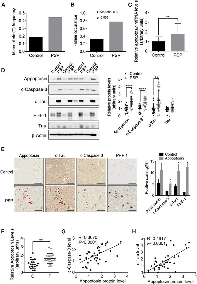
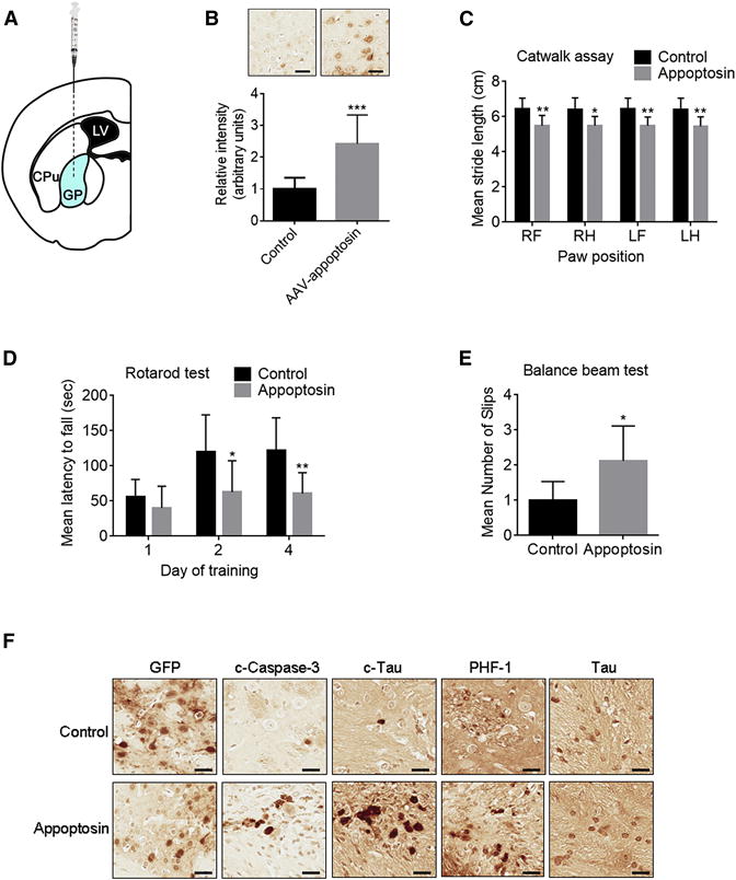
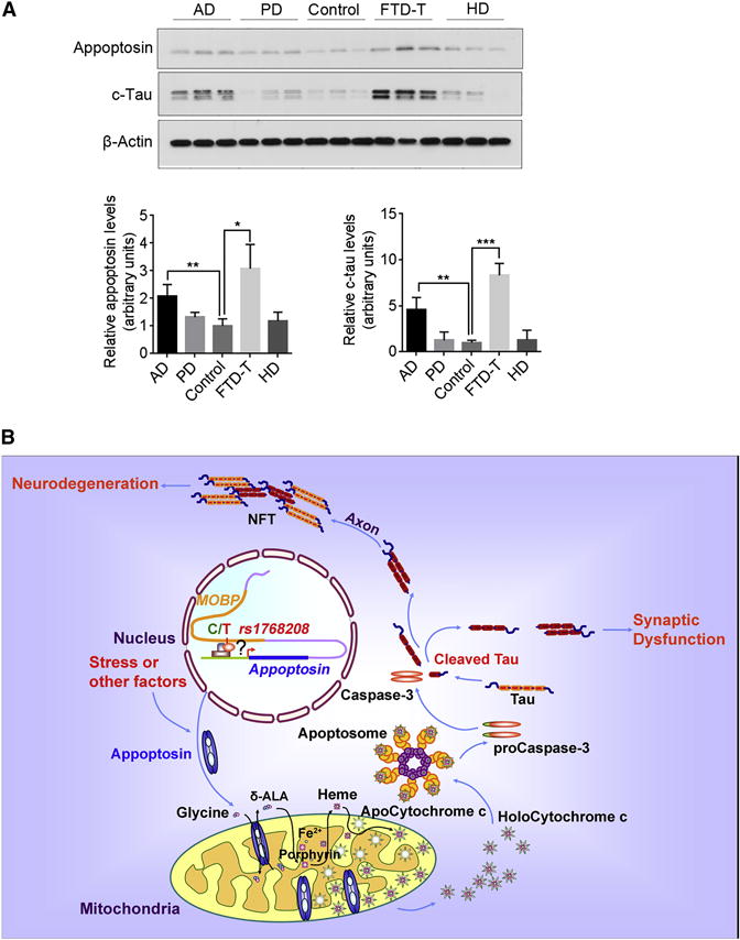
Progressive supranuclear palsy (PSP) is a movement disorder characterized by tau neuropathology where the underlying mechanism is unknown. An SNP (rs1768208 C/T) has been identified as a strong risk factor for PSP. Here, we identified a much higher T-allele occurrence and increased levels of the pro-apoptotic protein appoptosin in PSP patients. Elevations in appoptosin correlate with activated caspase-3 and caspase-cleaved tau levels. Appoptosin overexpression increased caspase-mediated tau cleavage, tau aggregation, and synaptic dysfunction, whereas appoptosin deficiency reduced tau cleavage and aggregation. Appoptosin transduction impaired multiple motor functions and exacerbated neuropathology in tau-transgenic mice in a manner dependent on caspase-3 and tau. Increased appoptosin and caspase-3-cleaved tau were also observed in brain samples of patients with Alzheimer's disease and frontotemporal dementia with tau inclusions. Our findings reveal a novel role for appoptosin in neurological disorders with tau neuropathology, linking caspase-3-mediated tau cleavage to synaptic dysfunction and behavioral/motor defects.


Figures

Figure 1
Figure 1. Elevated Appoptosin Levels Associate with Higher T-Allele Occurrence at SNP rs1768208 and Correlate with Increased Caspase-3 Activation and Tau Cleavage in PSP Brains
(A and B) The frequency and occurrence of the minor T- allele at SNP rs1768208 (C/T) in control and PSP patients. T occurrence: number of cases with T-allele/number of total cases (Control, n=22; PSP, n=26). Odds ratio is calculated with R statistical software. (C) Relative appoptosin mRNA levels in control and PSP brain samples (Control, n=15; PSP, n=23). (D) Western blot analysis of appoptosin, c-caspase-3, c-tau, PHF-1 and tau levels in the frontal cortex of PSP and control individuals (Control, n=15; PSP, n=23). (E) Immunohistochemistry analysis of appoptosin, c-caspase-3 and c-tau and PHF-1 in the cortex of PSP and control individuals (n=4). Scale bar, 100 μm. Red arrows indicate some of the cells double stained appoptosin and c-tau in adjacent sections. (F) Relative appoptosin protein levels in SNP rs1768208 T allele carriers and non-T allele (C) carriers (n=19 per group). (G and H) The correlation between c-caspase-3 and appoptosin (G), and between c-tau and appoptosin (H) protein levels (Regression analysis, n=38). Data represent mean ± SD. *P < 0.05, **P < 0.01, ****P < 0.0001 by nonparametric t-test. See also Figure S1.
Figure 2
Figure 2. Appoptosin Regulates Tau Cleavage through Caspase-3 Activation
(A) Change in c-caspase-3, c-tau and tau levels in neuronal cultures following appoptosin transduction as determined by western blot (n=3 per group, two-tailed Student’s t-test). (B) Western blot analysis of c-tau, c-caspase3, tau and PSD95 in cortical neuronal cultures from appoptosin knockout mouse embryos or littermate controls (n=3 per group, two-tailed Student’s t-test). (C) Change in c-tau and cleaved-PARP (c-PARP) levels in neuronal cultures after appoptosin overexpression and exposure to DMSO (−), caspase-3 inhibitor (C3I), or caspase-9 inhibitor (C9I) (n=3 per group, repeated-measures one-way ANOVA with Dunnett’s post hoc analysis). Data represent mean ± SD. *P < 0.05, **P < 0.01, and #P < 0.05. See also Figure S2.
Figure 3
Figure 3. Overexpression of Appoptosin Reduces Tau Association with Microtubules and Promotes Tau Cleavage
(A) Change in tau found in microtubule-bound and unbound fractions following appoptosin overexpression and treatment with C3I (n=3 per group, repeated-measures One-way ANOVA with Dunnett’s post hoc analysis). (B) Quantification of SDS-insoluble tau aggregates in neuronal cultures following appoptosin transduction as determined by filter/trap assays. SDS-soluble tau was used as a control (n=3 per group, two-tailed Student’s t-test). (C) Staining of c-tau and MAP2 in neurons transduced with AAV-appoptosin or AAV-GFP. Green color represents GFP without staining, which was used to identify transduced neurons (n=15 per group, two-tailed Student’s t-test). Scale bar, 20 μm. (D) c-Tau puncta in dendrites of appoptosin KO or littermate control neurons were detected by immunocytochemistry (n=15 per group, two-tailed Student’s t-test). Scale bar, 20 μm. Data represent mean ± SD. *P < 0.05, **P < 0.01, and #P < 0.05. Data represent mean ± SD.
Figure 4
Figure 4. Overexpression of Appoptosin Promotes c-Tau Distribution to Post-Synaptic Compartments, Disrupts Synaptic Structure and Depletes Cell Surface Glutamate Receptors
(A) Distribution of c-tau, c-caspase-3, and total tau in postnuclear (S1), synaptosomal (Syn) and PSD fractions after appoptosin overexpression (n=3 per group, two-tailed Student’s t-test). (B and C) DIV (days in vitro) 14 neurons were transfected or transduced with AAV-appoptosin. Numbers of dendritic spines (B) and Synapsin-I/PSD95 double-positive puncta (C) in neurons with appoptosin overexpression and caspase-3 inhibition (n=15 per group, one-way ANOVA with Tukey’s post hoc analysis). Scale bar, 10 μm. (D) Cell surface levels of NR1, NR2A, NR2B, GluR1, GluR2 and Na+/K+ATPase (as a control) in neurons with appoptosin overexpression, as determined by cell surface biotinylation assays (n=3 per group, two-tailed Student’s t test). Data represent mean ± SD. *P < 0.05, **P < 0.01, ***P < 0.001, ****P < 0.0001, and #P < 0.05. See also Figure S3.
Figure 5
Figure 5. Overexpression of Appopotosin Causes PSP-like Motor Deficits and Induces Tau Pathology
(A) Schematic workflow of stereotaxic AAV injection into the globus pallidus of JNPL3 mice. LV, lateral ventricle; CPu, caudate putamen; GP: globus pallidus. (B–F) Behavioral and histological analysis of JNPL3 mice injected with AAV-appoptosin or AAV-GFP (control). (B) Confirmation of appoptosin overexpression within the injected area (n=8 per group, two-tailed Student’s t test). Scale bar, 30 μm. (C) Catwalk analysis: comparison of stride length of each limb (RF, right front; RH, right hind; LF, left front; LH, left hind) of experimental mice (n=8 per group, two-way ANOVA with Sidak’s post hoc analysis). (D) Accelerating rotarod test: latency of experimental mice on the rotor at days after training as indicated (n=8 per group, two-way ANOVA with Sidak’s post hoc analysis). (E) Number of slips observed in experimental mice in balance beam tests (n=8 per group, two-tailed Student’s t test). (F) The expression of GFP, c-caspase-3, c-tau, PHF-1 tau, and tau in the injected area in experimental mice. n=8, Scale bar, 30 μm. Data represent mean ± SD. *P < 0.05, **P < 0.01, and ***P < 0.001. See also Figure S4.
Figure 6
Figure 6. Caspase Inhibition and Tau Knockout Reduces Detrimental Effect Caused by Appoptosin Overexpression
(A, B and C) Behavioral and histological analysis of JNPL3 mice injected with AAV-appoptosin or AAV-GFP (control), and treated with or without caspase-3 inhibitor. (A) Comparison of stride length of each limb (RF, right front; RH, right hind; LF, left front; LH, left hind) of experimental mice (n=11 per group, two-way ANOVA with Tukey’s post hoc analysis). (B) Immunostaining to show expression of c-Tau, PHF1, NeuN, GFAP, and MAP2 in the infection area. (C) Quantification of immunostaining from JNPL3 mice (n=8 per group, one-way ANOVA with Dunnett’s post hoc analysis) (D and E) Behavioral and histological analysis of wild type (WT) mice and tau knockout (KO) mice injected with AAV-appoptosin or AAV-GFP. (D) Number of errors made by experimental mice during the round beam test (n=12 for WT and Tau KO mice, n=9 for WT-appoptosin mice, n=11 for Tau KO-appoptosin mice, two-way ANOVA with Sidak’s post hoc analysis). (E) Expression of NeuN, GFAP, MAP2 and c-caspase-3 in the injection area (n=11 for WT and Tau KO-appoptosin mice, n=9 for WT-appoptosin mice, n=10 for Tau KO mice, two-way ANOVA with Sidak’s post hoc analysis). Scale bar, 50 μm. Data represent mean ± SD. *P < 0.05, **P < 0.01, and ***P < 0.001. See also Figure S4G.
Figure 7
Figure 7. Appoptosin Is Upregulated in Tauopathic Disorders, and Drives Pathogenesis through Caspase Activation, Tau Cleavage and Aggregation
(A) Analysis of appoptosin and c-tau expression in the frontal cortex of AD, PD, control, FTD-T and HD individuals (AD, n=8; PD, n=3; control, n=3; FTD-T, n=3; HD, n=3). Data represent mean ± SD. *P < 0.05, **P < 0.01, ***P < 0.001, two-tailed Student’s t-test. (B) Appoptosin potentially drives PSP pathogenesis through caspase-3 mediated tau cleavage and NFT formation. Appoptosin expression is increased in PSP individuals through a C/T polymorphism at the SNP rs1768208 or stress. Increased appoptosin protein increases heme synthesis, and thus activates the intrinsic caspase pathway and caspase-mediated tau cleavage. Cleaved tau (c-tau) is prone to aggregate to form neurofibrillary tangles (NFT), which is deleterious to neurons. c-Tau also can be mis-sorted into the postsynaptic compartment to trigger synaptic dysfunction. Together with caspase activation, these events eventually lead to tau pathology and neurodegeneration in PSP.

References

    1. Baker M, Litvan I, Houlden H, Adamson J, Dickson D, Perez-Tur J, Hardy J, Lynch T, Bigio E, Hutton M. Association of an extended haplotype in the tau gene with progressive supranuclear palsy. Hum Mol Genet. 1999;8:711–715. - PubMed
    1. Ballatore C, Lee VM, Trojanowski JQ. Tau-mediated neurodegeneration in Alzheimer’s disease and related disorders. Nature Rev Neurosci. 2007;8:663–672. - PubMed
    1. Blin N, Stafford DW. A general method for isolation of high molecular weight DNA from eukaryotes. Nucleic Acids Res. 1976;3:2303–2308. - PMC - PubMed
    1. Borroni B, Agosti C, Magnani E, Di Luca M, Padovani A. Genetic bases of Progressive Supranuclear Palsy: the MAPT tau disease. Curr Med Chem. 2011;18:2655–2660. - PubMed
    1. Conrad C, Andreadis A, Trojanowski JQ, Dickson DW, Kang D, Chen X, Wiederholt W, Hansen L, Masliah E, Thal LJ, et al. Genetic evidence for the involvement of tau in progressive supranuclear palsy. Ann Neurol. 1997;41:277–281. - PubMed

Publication types

MeSH terms