Review
. 2015 Dec;79(4):387-401.
doi: 10.1128/MMBR.00027-15.

Long-Term Relationships: the Complicated Interplay between the Host and the Developmental Stages of Toxoplasma gondii during Acute and Chronic Infections

Affiliations
Review

Long-Term Relationships: the Complicated Interplay between the Host and the Developmental Stages of Toxoplasma gondii during Acute and Chronic Infections

Kelly J Pittman et al. Microbiol Mol Biol Rev. 2015 Dec.

Abstract

Toxoplasma gondii represents one of the most common parasitic infections in the world. The asexual cycle can occur within any warm-blooded animal, but the sexual cycle is restricted to the feline intestinal epithelium. T. gondii is acquired through consumption of tissue cysts in undercooked meat as well as food and water contaminated with oocysts. Once ingested, it differentiates into a rapidly replicating asexual form and disseminates throughout the body during acute infection. After stimulation of the host immune response, T. gondii differentiates into a slow-growing, asexual cyst form that is the hallmark of chronic infection. One-third of the human population is chronically infected with T. gondii cysts, which can reactivate and are especially dangerous to individuals with reduced immune surveillance. Serious complications can also occur in healthy individuals if infected with certain T. gondii strains or if infection is acquired congenitally. No drugs are available to clear the cyst form during the chronic stages of infection. This therapeutic gap is due in part to an incomplete understanding of both host and pathogen responses during the progression of T. gondii infection. While many individual aspects of T. gondii infection are well understood, viewing the interconnections between host and parasite during acute and chronic infection may lead to better approaches for future treatment. The aim of this review is to provide an overview of what is known and unknown about the complex relationship between the host and parasite during the progression of T. gondii infection, with the ultimate goal of bridging these events.


Figures

FIG 1
FIG 1
Sexual reproduction of T. gondii. Felines, the definitive host, are most often infected with T. gondii (depicted above in green) through ingestion of an infected host. Once ingested, T. gondii penetrates the epithelial cells of the small intestine and differentiates into tachyzoite and schizont stages. The asexual tachyzoites divide and disseminate throughout the feline. The schizont will remain within the intestinal epithelium and has 5 distinct stages, identified as types A to E. They are classified based on their mode of division, the time postinfection they are observed, and their structural components. Type E schizonts give rise to merozoites, which differentiate into gametes. Gametes can be found throughout the small intestine as soon as 3 days postinfection and can last for a few weeks postinoculation with tissue cysts. Males (microgametes) fertilize females (macrogametes) to produce oocysts. After fertilization occurs, the oocyst wall forms around the parasite. Sporulation of oocysts occurs 1 to 5 days after being excreted in cat feces. Once sporulation occurs, oocysts are infectious for an extended period of time, depending on environmental conditions.
FIG 2
FIG 2
Ingestion of T. gondii. (A) Inoculation with T. gondii occurs from ingestion of undercooked meat containing encysted bradyzoites or food contaminated with sporozoites within oocysts. (B) Bradyzoites or sporozoites are released into the lumen of the small intestine, where they invade intestinal enterocytes. (C to E) Parasites differentiate into the tachyzoite form (C), where they rapidly replicate (D) and egress from the cell (E). (F) Parasites begin invading cells of the lamina propria and encounter resident immune cells. (G) Infected resident immune cells produce chemokines to trigger migration and effector functions of circulating immune cells.
FIG 3
FIG 3
Innate immune response. (A) Chemokines secreted from cells at the site of infection, such as MCP-1 and MIP-2, trigger migration of circulating neutrophils and inflammatory monocytes to the site of infection. (B) Inflammatory monocytes differentiate into dendritic cells and macrophages, where they, along with neutrophils, generate IL-12 in response to infection. (C) IL-12 stimulates NK cells and CD4+ and CD8+ T cells to produce IFN-γ.
FIG 4
FIG 4
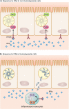
Cellular response to IFN-γ. (A) A variety of host cells respond to T. gondii when stimulated with IFN-γ by upregulating genes with antimicrobial functions. Two large protein families that are IFN-γ inducible are guanylate-binding proteins (GBPs) and immunity-related GTPases (IRGs). GBPs and IRGs accumulate at the parasitophorous vacuole, which is formed upon entry of the parasite, and aid in restriction of growth and destruction of T. gondii (depicted in light gray). (B) Different IFN-γ-inducible mechanisms are used in immune cells, such as production of ROS and RNS. Reactive oxygen and nitrogen species are toxic and function to create unfavorable and unstable environments for intracellular parasites.
FIG 5
FIG 5
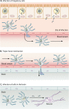
Infection of immunoprivileged sites. (A) Migratory immune cells arrive at the site of infection to aid in the clearance of T. gondii. Some immune cells become infected, leave the site of infection, and travel to other tissue carrying live parasites. (B) T. gondii is able to cross barriers, such as the blood-brain barrier, into immune-privileged sites by invading migratory immune cells; this is referred to as the Trojan horse mechanism. (C) Once in the brain, or another immunoprivileged site, T. gondii egresses from the cell and invades resident cells. In the central nervous system, T. gondii tachyzoites will differentiate to the bradyzoite stage, form a cyst wall, and establish a chronic infection.

References

    1. Lindsay DS, Dubey JP. 2014. Toxoplasmosis in wild and domestic animals, p 194-209. In Weiss LM, Kim K (ed), Toxoplasma gondii: the model apicomplexan, 2nd ed Elsevier, Amsterdam, The Netherlands.
    1. Frenkel JK, Dubey JP, Miller NL. 1970. Toxoplasma gondii in cats: fecal stages identified as coccidian oocysts. Science 167:893–896. doi:10.1126/science.167.3919.893. - DOI - PubMed
    1. Dubey JP, Lindsay DS, Speer CA. 1998. Structures of Toxoplasma gondii tachyzoites, bradyzoites, and sporozoites and biology and development of tissue cysts. Clin Microbiol Rev 11:267–299. - PMC - PubMed
    1. Munoz-Zanzi CA, Fry P, Lesina B, Hill D. 2010. Toxoplasma gondii oocyst-specific antibodies and source of infection. Emerg Infect Dis 16:1591–1593. doi:10.3201/eid1610.091674. - DOI - PMC - PubMed
    1. Cook AJ, Gilbert RE, Buffolano W, Zufferey J, Petersen E, Jenum PA, Foulon W, Semprini AE, Dunn DT. 2000. Sources of toxoplasma infection in pregnant women: European multicentre case-control study. European Research Network on Congenital Toxoplasmosis. BMJ 321:142–147. doi:10.1136/bmj.321.7254.42. - DOI - PMC - PubMed

Publication types