

Alterations in the rostral ventromedial medulla after the selective ablation of μ-opioid receptor expressing neurons
- PMID: 26335909
- PMCID: PMC4829402
- DOI: 10.1097/j.pain.0000000000000344
Alterations in the rostral ventromedial medulla after the selective ablation of μ-opioid receptor expressing neurons
Abstract
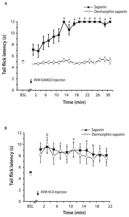
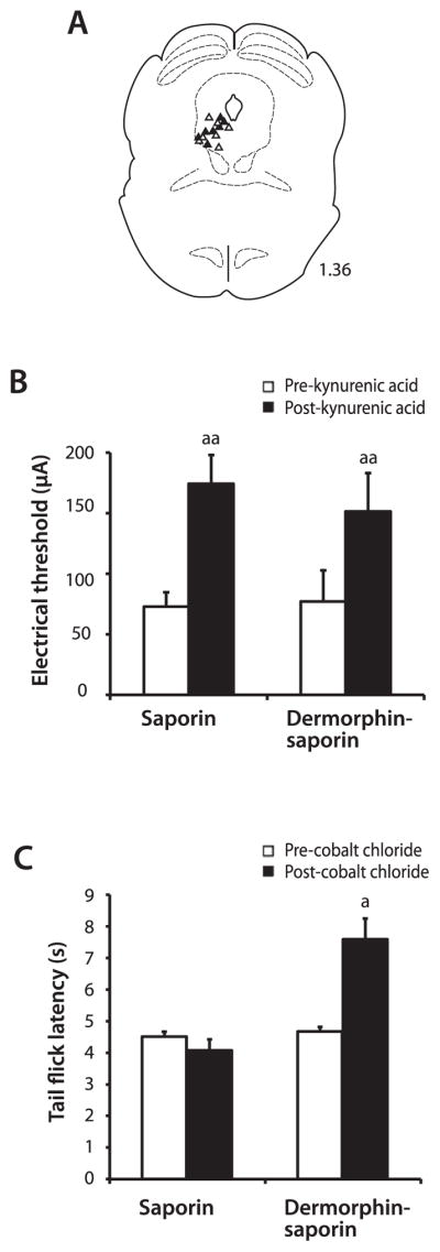
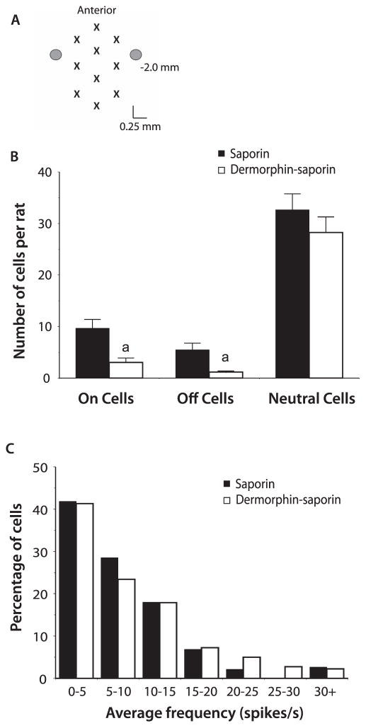
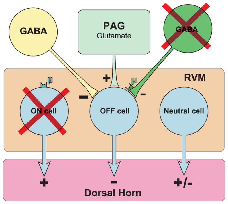
The rostral ventromedial medulla (RVM) exerts both inhibitory and excitatory controls over nociceptive neurons in the spinal cord and medullary dorsal horn. Selective ablation of mu-opioid receptor (MOR)-expressing neurons in the RVM using saporin conjugated to the MOR agonist dermorphin-saporin (derm-sap) attenuates stress and injury-induced behavioral hypersensitivity, yet the effect of RVM derm-sap on the functional integrity of the descending inhibitory system and the properties of RVM neurons remain unknown. Three classes of RVM neurons (on-cells, off-cells, and neutral cells) have been described with distinct responses to noxious stimuli and MOR agonists. Using single unit recording in lightly anesthetized rats, RVM neurons were characterized after microinjections of derm-sap or saporin. Derm-sap treatment resulted in a reduction in on-cells and off-cells when compared to saporin controls (P < 0.05). The number of neutral cells remained unchanged. After derm-sap treatment, RVM microinjections of the glutamate receptor agonist homocysteic acid increased tail-flick latencies, whereas the MOR agonist DAMGO had no effect. Furthermore, electrical stimulation of the periaqueductal gray produced analgesia in both derm-sap and saporin controls with similar thresholds. Microinjection of kynurenic acid, a glutamate receptor antagonist, into the RVM disrupted periaqueductal gray stimulation-produced analgesia in both saporin-treated and derm-sap-treated rats. These results indicate that MOR-expressing neurons in the RVM are not required for analgesia produced by either direct or indirect activation of neurons in the RVM.
Conflict of interest statement
The authors have no conflicts of interest to declare.
Figures

References
-
- Bederson JB, Fields HL, Barbaro NM. Hyperalgesia during naloxone-precipitated withdrawal from morphine is associated with increased on-cell activity in the rostral ventromedial medulla. Somatosens Mot Res. 1990;7:185–203. - PubMed
-
- Bee LA, Dickenson AH. Descending facilitation from the brainstem determines behavioural and neuronal hypersensitivity following nerve injury and efficacy of pregabalin. PAIN. 2008;140:209–23. - PubMed
-
- Behbehani MM, Fields HL. Evidence that an excitatory connection between the periaqueductal gray and nucleus raphe magnus mediates stimulation produced analgesia. Brain Res. 1979;170:85–93. - PubMed
-
- Beitz A. Relationship of glutamate and aspartate to the periaqueductal gray-raphe magnus projection: analysis using immunocytochemistry and microdialysis. J Histochem Cytochem. 1990;38:1755–65. - PubMed
MeSH terms
Substances
Grants and funding
LinkOut - more resources
Full Text Sources
Other Literature Sources
Medical
Research Materials
Miscellaneous
