Review
. 2015 Oct;95(4):1205-40.
doi: 10.1152/physrev.00037.2014.

Membrane Repair: Mechanisms and Pathophysiology

Affiliations
Review

Membrane Repair: Mechanisms and Pathophysiology

Sandra T Cooper et al. Physiol Rev. 2015 Oct.

Abstract

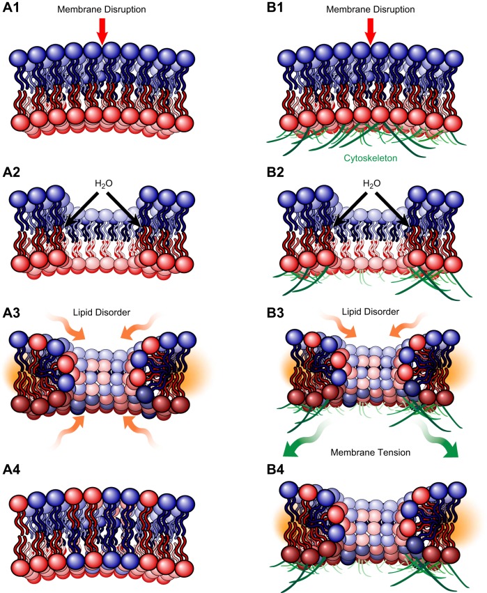
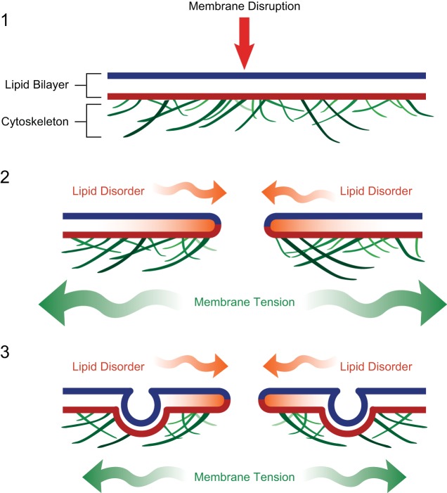
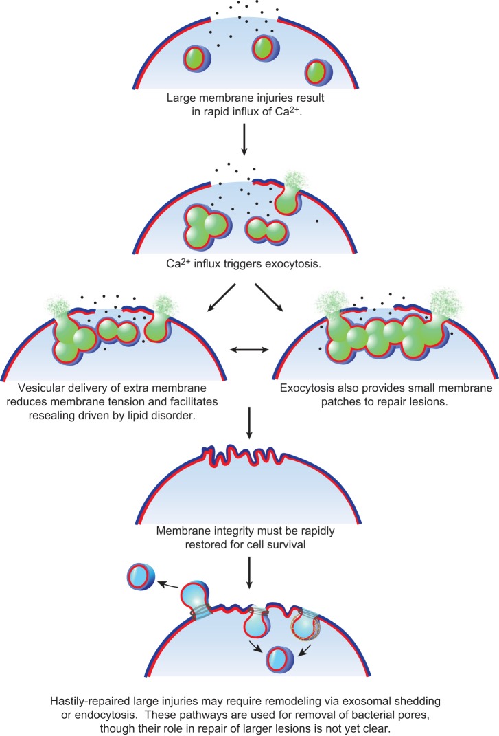
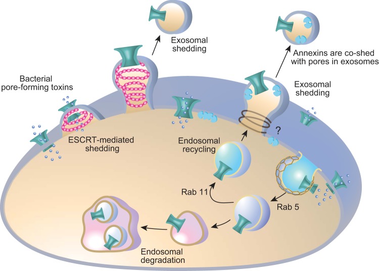
Eukaryotic cells have been confronted throughout their evolution with potentially lethal plasma membrane injuries, including those caused by osmotic stress, by infection from bacterial toxins and parasites, and by mechanical and ischemic stress. The wounded cell can survive if a rapid repair response is mounted that restores boundary integrity. Calcium has been identified as the key trigger to activate an effective membrane repair response that utilizes exocytosis and endocytosis to repair a membrane tear, or remove a membrane pore. We here review what is known about the cellular and molecular mechanisms of membrane repair, with particular emphasis on the relevance of repair as it relates to disease pathologies. Collective evidence reveals membrane repair employs primitive yet robust molecular machinery, such as vesicle fusion and contractile rings, processes evolutionarily honed for simplicity and success. Yet to be fully understood is whether core membrane repair machinery exists in all cells, or whether evolutionary adaptation has resulted in multiple compensatory repair pathways that specialize in different tissues and cells within our body.


Figures

Figure 1.
Figure 1.
Spontaneous resealing of plasma membrane injuries in the nanometer range is opposed by the forces of the underlying membrane cytoskeleton. For an injury to a phospholipid bilayer alone (A1-4), the lipid disorder present on the curved edges of the disruption provides the driving force to spontaneously reseal the injury and is a function of disruption diameter squared. However, if the injured phospholipid bilayer is tethered to underlying cytoskeleton (B1-4), the membrane tension from adhesion to the cytoskeleton confers an opposing force for resealing, a function of disruption diameter cubed, and prevents spontaneous repair of membrane disruptions that exceed diameters in the nanometer range (106).
Figure 2.
Figure 2.
Calcium-activated exocytosis reduces membrane tension and promotes spontaneous repair driven by lipid disorder for injuries hundreds of nanometers in diameter. With larger injuries, the opposing force of membrane tension exceeds the resealing forces of lipid disorder at the edges of the disruption, negating the driving forces of spontaneous membrane resealing. Eukaryotic cells have been shown to utilize calcium-activated exocytosis to reduce membrane tension and promote repair via lipid-disorder driven attractions. The reduction in membrane tension is likely due directly to the addition of phospholipids to reduced lipid packing, as well as due in part to the cytoskeletal remodeling associated with vesicular transport at the plasma membrane.
Figure 3.
Figure 3.
Very large plasma membrane disruptions (micron diameter) require membrane patching. The calcium influx of a membrane injury activates vesicular exocytosis and homo- and heterotypic fusion of cytoplasmic vesicles. Exocytic fusion reduces membrane tension, and vesicle-vesicle fusion events provide a patch as a replacement for the membrane barrier missing at the disruption site. The membrane patch may serve only temporarily as a surface barrier replacement that is subsequently remodeled and removed via exocytic and/or endocytic machinery.
Figure 4.
Figure 4.
Survival from bacterial pore-forming toxins utilizes both exocytic and endocytic responses. Bacterial pore-forming toxins oligomerize and insert in the plasma membrane of target cells forming a diffusible pore. Evidence suggests these pores are removed both by endosomal degradative pathways (123, 164, 280) and exosomal shedding (14, 118, 136). Shed microvesicles containing streptolysin-O have been shown to also contain annexins A1 and A6 (219). In C. elegans, Rab 5 endocytic and Rab 11 recycling pathways are implicated in pore removal (164). ESCRT machinery (pink helix) has been implicated in exosomal shedding (129), although other exosomal machinery may also be involved (indicated by a question mark).
Figure 5.
Figure 5.
What might membrane injury to muscle fibers look like? Muscle fibers have a complex plasma membrane network with a repeating register of deep plasma membrane invaginations called the t-tubule network. Muscle fibers are subject to huge variations in membrane tension, due to their contractile activity. Repeated eccentric exercise in healthy subjects (i.e., stepping down for 20 min) is known to induce damage so severe that muscle fibers degenerate over the following days and weeks (91, 131, 199). Patients with muscular dystrophy are more susceptible to injury from eccentric stretch (216), with studies in mouse models suggesting susceptibility to injury can escalate with multiple insults (53). One model explaining membrane injury in dystrophin-deficient muscle fibers proposes that an initial injury causes a local influx of calcium and a local region of hypercontraction. These shortened sarcomeres induce a concomitant lengthening of adjacent sarcomeres and increased lateral strain to the plasma membrane. Subsequent insult(s) of eccentric stretch result in a more severe wound and global hypercontraction, producing fiber retraction within the muscle bundle (53). As muscle fibers have strong interfiber connections, muscle injuries may manifest both as shearing of the membrane from increased membrane tension and strain, as well as ripping of plasma membrane regions from fiber retraction or hypercontraction.
Figure 6.
Figure 6.
Schematic representation of the structural features of the protein families implicated in membrane repair.
Figure 7.
Figure 7.
A cartoon depicting the potential role of dysferlin-mediated vesicle fusion in membrane repair. Membrane injury causes a local influx of calcium and activation of calpains. MG53 (40) shows diffuse enrichment at injury sites within 2 s of membrane injury in a calcium-independent manner (150). The signal to activate recruitment of MG53 to injury sites is not clear, but may relate to its role as a ubiquitin ligase to target substrate(s) damaged as a consequence of the membrane injury. Dysferlin is not detected at injury sites until 10 s postinjury, a delay we attribute to an intermediary step involving calpain cleavage. Activated calpains cleave dysferlin within a motif specifically encoded by alternately spliced exon 40a (230). As dysferlin may only be detected at injury sites with antibodies recognizing COOH-terminal epitopes, and not several antibodies to NH2-terminal or central domains (150), data suggest the COOH-terminal cleaved fragment termed mini-dysferlinC72 is the form specifically recruited to injury sites. It remains uncertain whether full-length dysferlin is also present at injury sites but is structurally precluded from antibody labeling and whether the dysferlin recruited for membrane repair is derived from plasma membrane or intracellular membrane compartments. Super-resolution 3-dimensional structured illumination microscopy (3D-SIM) reveals dysferlin-laden vesicles undergo calcium-dependent integration at injury sites (150). At 10 s postinjury, dysferlin and MG53 form a lattice that intensely labels the edges of the lesions. Lesions expand and are filled in by a dysferlin and MG53 lattice. At 90 s postinjury, filled lesions are characterized by a dominant arc of dysferlin and MG53 labeling we believe represents the original edges of the lesion that have been drawn together by cytoskeletal motors for resealing. [3D-SIM images from Lek et al. (150), with permission from The Journal of Neuroscience.]

References

    1. Abrami L, Fivaz M, Glauser PE, Parton RG, van der Goot FG. A pore-forming toxin interacts with a GPI-anchored protein and causes vacuolation of the endoplasmic reticulum. J Cell Biol 140: 525–540, 1998. - PMC - PubMed
    1. Abreu-Blanco MT, Verboon JM, Parkhurst SM. Cell wound repair in Drosophila occurs through three distinct phases of membrane and cytoskeletal remodeling. J Cell Biol 193: 455–464, 2011. - PMC - PubMed
    1. Achanzar WE, Ward S. A nematode gene required for sperm vesicle fusion. J Cell Sci 110: 1073–1081, 1997. - PubMed
    1. Adibhatla RM, Hatcher JF. Lipid oxidation and peroxidation in CNS health and disease: from molecular mechanisms to therapeutic opportunities. Antioxidants Redox Signal 12: 125–169, 2010. - PubMed
    1. Allen DG. Eccentric muscle damage: mechanisms of early reduction of force. Acta Physiol Scand 171: 311–319, 2001. - PubMed

Publication types