. 2015 Oct 16;290(42):25745-55.
doi: 10.1074/jbc.M115.669598. Epub 2015 Sep 2.

Competing Lipid-Protein and Protein-Protein Interactions Determine Clustering and Gating Patterns in the Potassium Channel from Streptomyces lividans (KcsA)

Affiliations

Competing Lipid-Protein and Protein-Protein Interactions Determine Clustering and Gating Patterns in the Potassium Channel from Streptomyces lividans (KcsA)

M Luisa Molina et al. J Biol Chem. .

Abstract

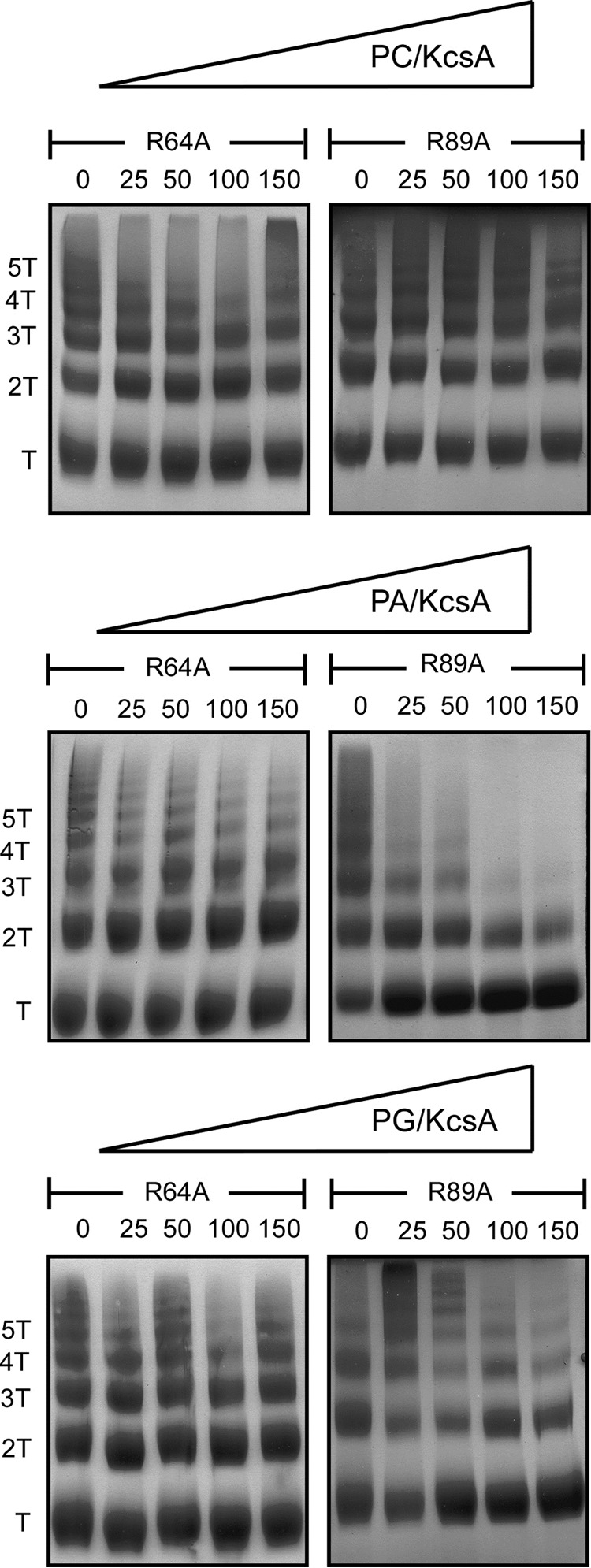
There is increasing evidence to support the notion that membrane proteins, instead of being isolated components floating in a fluid lipid environment, can be assembled into supramolecular complexes that take part in a variety of cooperative cellular functions. The interplay between lipid-protein and protein-protein interactions is expected to be a determinant factor in the assembly and dynamics of such membrane complexes. Here we report on a role of anionic phospholipids in determining the extent of clustering of KcsA, a model potassium channel. Assembly/disassembly of channel clusters occurs, at least partly, as a consequence of competing lipid-protein and protein-protein interactions at nonannular lipid binding sites on the channel surface and brings about profound changes in the gating properties of the channel. Our results suggest that these latter effects of anionic lipids are mediated via the Trp(67)-Glu(71)-Asp(80) inactivation triad within the channel structure and its bearing on the selectivity filter.

Keywords: Ion channel inactivation; Supramolecular assembly of ion channels; coupled channel gating; gating; lipid-protein interaction; membrane protein clusters; membrane protein nonannular sites; molecular modeling; potassium channel; protein-protein interaction.


Figures

FIGURE 1.
FIGURE 1.
Lipid-protein and protein-protein interactions at nonannular lipid binding sites. A shows a top view (normal to the membrane plane) of an extracellular portion comprising amino acid residues 62–101 (roughly indicated by the box on the small diagram to the right of A) in the crystallographic structure of KcsA (Protein Data Bank code 1K4C) defining one of the four intersubunit crevices acting as nonannular lipid binding sites. A DPPA molecule (atoms represented as spheres) has been drawn bound to such a site, using as a scaffold the partial lipid structure appearing in the protein crystal (see “Materials and Methods”). Adjacent protein subunits defining the nonannular site are colored in light brown and green. The one-letter codes and numbering in the KcsA sequence for the main amino acid residues involved in the interactions have been included. Hydrogen bonds are indicated by red dashed lines. It should be noted that the Trp67–Glu71–Asp80 inactivation triad is fully configured and that the lipid polar head interacts primarily with Arg64 and Arg89. B shows the results from computer docking two KcsA channels in an attempt to model a cluster. In this case, the KcsA sequence was previously modeled on the basis of homology onto the open structure of MthK (see “Materials and Methods”). The nonannular binding sites, flanked by the pore helix and M2 in each channel, interact now with the M2 from the adjacent channel (channel 2; subunits colored in light blue and pink), whereas the inactivation triad has been disrupted. C shows the results of modeling the two KcsA channel cluster based on the x-ray structure of the potassium transporter TrkH, which already crystallizes as a dimer (see “Materials and Methods”). Notice that in this case, the nonannular binding site interacts with the pore helix from the adjacent channel, but the effect of such interaction on the disruption of the inactivation triad is identical.
FIGURE 2.
FIGURE 2.
Blue Native PAGE analysis of phospholipid effects on the disassembly of KcsA clusters in mixed micelles. Mixed micelles containing KcsA and either PC, PE, PG, and PA at the indicated phospholipid to KcsA molar ratios were prepared and analyzed by BN-PAGE. Representative gels for each condition and the results from densitometry are expressed as percentages of T (closed squares) and nT (open squares) bands relative to the sum of all the bands present within each gel lane are shown. T stands for the KcsA tetramer, whereas nT refers to the sum of 2T, 3T, 4T, and 5T cluster species having 2, 3, 4, and 5 times the molecular weight of the KcsA tetramer. The values represent the averages ± S.E. of five different gels from each condition.
FIGURE 3.
FIGURE 3.
KcsA clusters in supported bilayers. Representative fluorescence microscopy images of a confocal cross-section (parallel to the bilayer plane) of SLBs containing Alexa 647-labeled KcsA. A, SLBs made from a DOPC:DOPG mixture at a 95:5 molar ratio. B, SLBs of pure DOPG. Large and highly fluorescent array-like protein complexes of variable sizes are seen in both cases. C, size distribution of KcsA clusters in the two SLBs samples from above. Bars represent the number of clusters observed per image in the pure DOPG (filled) and in the DOPC:DOPG (open) samples. The values are the averages ± S.E. of four to nine images from each of three different SLB samples prepared in each condition.
FIGURE 4.
FIGURE 4.
Effects of anionic phospholipids on the gating patterns of KcsA reconstituted into giant liposomes. A and B show representative voltage ramps (−200 to 200 mV from a 0 mV holding potential, 133 mV/s) showing typical LOP and HOP activity patterns of KcsA obtained by patch clamping excised, inside-out patches from giant liposomes containing KcsA reconstituted into 5 or 25% of anionic lipid (PA or PG; see “Materials and Methods”). In this figure, as well as in Fig. 6, dashed lines indicate the closed channel states, and thin continuous lines indicate zero current level. Channel openings appear as upward (at positive voltages) or downward (at negative voltages) deflections over the closed state line. C shows the percentage of patches showing HOP patterns in each of the above groups. Giant liposomes containing only 5% (black columns) of either PA or PG in the PC/cholesterol matrix showed 58.3% (n = 14 from 24 patches) or 75.0% (n = 45 from 60 patches) of patches exhibiting HOP patterns of activity, respectively, whereas giant liposomes containing 25% (gray columns) of either PA or PG in the lipid matrix show 17.9% (n = 7 from 39 patches) or 38.5% (n = 20 from 52 patches) HOP patterns of activity, respectively. To compare the occurrence of a characteristic of interest between two groups, we used the z test. Asterisks indicate significant differences (p < 0.05) in the occurrence of HOP pattern activity in samples containing different proportions of the same anionic lipid. The effect of anionic lipids on HOP activity occurrence was even more evident when comparing together the data of PA and PG for the two concentrations tested (see “Results” for details). The right-hand column in C, labeled ASO, indicates the percentage of HOP patterns found when reconstituting KcsA into giant liposomes made from asolectin lipids (45.7%; n = 75 from 164 patches). D, column histogram of the rectification index (see “Materials and Methods”) of the HOP pattern of KcsA channels reconstituted in 5 and 25% anionic lipids.
FIGURE 5.
FIGURE 5.
Blue Native PAGE analysis of the effects of phospholipids on the disassembly of clusters of KcsA arginine mutants in mixed micelles. Mixed micelles containing the indicated KcsA arginine mutants and either PC (top row), PA (middle row), or PG (bottom row), at the indicated phospholipid to KcsA molar ratios, were analyzed by BN-PAGE. Experimental conditions and details within the figure are as in Fig. 2.
FIGURE 6.
FIGURE 6.
Gating patterns observed in R64A and R89A KcsA mutants. A and D show representative voltage ramps illustrating the predominant activity patterns of the R64A (A; 17 of 25 patches, 68% of the cases) and the R89A (D; 30 of 41 patches, 73% of the cases) KcsA mutants, respectively, reconstituted into giant liposomes made from asolectin lipids. Experimental conditions and drawing details are as in Fig. 4. Notice that the R64A mutant shows HOP patterns of activity very similar to those seen in wild-type KcsA (12) and exhibits coupled gating, as illustrated in the continuous recording taken at +150 mV shown in B. None of these features are clearly present in the R89A mutant (D and E), which exhibits mainly uncoupled HOPs (see “Results”). C and F show single channel recordings taken also at +150 mV of the two mutant channels to illustrate that despite the above differences; the channel conductance and opening probability when analyzed at the single channel level are similar in both cases. G shows a column histogram of the rectification index of the HOP pattern of the wild-type KcsA, as well as those of the R64A and R89A mutant channels.
SCHEME 1
SCHEME 1

Similar articles

Cited by

References

    1. Doyle D. A., Morais Cabral J., Pfuetzner R. A., Kuo A., Gulbis J. M., Cohen S. L., Chait B. T., MacKinnon R. (1998) The structure of the potassium channel: molecular basis of K+ conduction and selectivity. Science 280, 69–77 - PubMed
    1. Zhou Y., Morais-Cabral J. H., Kaufman A., MacKinnon R. (2001) Chemistry of ion coordination and hydration revealed by a K+ channel-Fab complex at 2.0 A resolution. Nature 414, 43–48 - PubMed
    1. Uysal S., Vásquez V., Tereshko V., Esaki K., Fellouse F. A., Sidhu S. S., Koide S., Perozo E., Kossiakoff A. (2009) Crystal structure of full-length KcsA in its closed conformation. Proc. Natl. Acad. Sci. U.S.A. 106, 6644–6649 - PMC - PubMed
    1. Schrempf H., Schmidt O., Kümmerlen R., Hinnah S., Müller D., Betzler M., Steinkamp T., Wagner R. (1995) A prokaryotic potassium ion channel with two predicted transmembrane segments from Streptomyces lividans. EMBO J. 14, 5170–5178 - PMC - PubMed
    1. Meuser D., Splitt H., Wagner R., Schrempf H. (1999) Exploring the open pore of the potassium channel from Streptomyces lividans. FEBS Lett. 462, 447–452 - PubMed

Publication types

LinkOut - more resources