. 2015 Feb;31(1):33-7.
doi: 10.1016/j.joa.2014.05.005. Epub 2014 Jul 4.

Measurement of the ventriculoatrial interval from the coronary sinus during para-Hisian pacing may fail to distinguish ventriculoatrial nodal conduction from conduction over a septal accessory pathway

Affiliations

Measurement of the ventriculoatrial interval from the coronary sinus during para-Hisian pacing may fail to distinguish ventriculoatrial nodal conduction from conduction over a septal accessory pathway

Takafumi Iijima et al. J Arrhythm. 2015 Feb.

Abstract

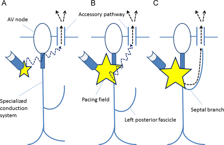
Background: Para-Hisian pacing (PHP) helps differentiate retrograde conduction over an accessory pathway (AP) from retrograde conduction over the atrioventricular (AV) node. This study examined a potential limitation of this technique, focusing on the measurement of the ventriculoatrial (V-A) interval from the coronary sinus (CS) during PHP.

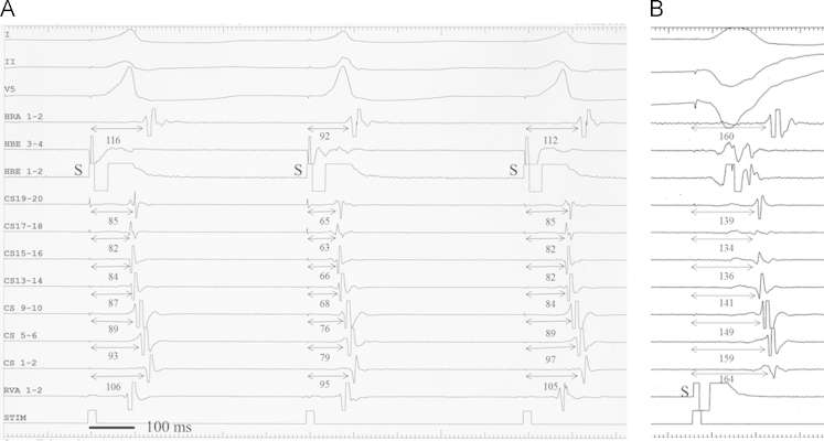
Methods: Our subjects were 9 patients undergoing electrophysiological studies before successful catheter ablation of a posteroseptal AP. During PHP, retrograde conduction occurred over an AP when the pacing stimulus to atrium (S-A) interval recorded near the AP remained unchanged whether the His bundle (HB) was captured or not (pattern 1), or when a loss of HB capture was associated with an increase in the S-A interval and no change in the V-A interval near the AP (pattern 2).

Results: Patterns 1 and 2 were observed in 5 (56%) and 2 (22%) patients, respectively. However, in the remaining 2 patients (22%), loss of HB capture during PHP was associated with an increase in the S-A interval (as in pattern 2), whereas the V-A interval near the AP could not be measured because no ventricular electrogram was visible on the CS recording (pattern 3); therefore, the presence of AP could not be confirmed by PHP. In patterns 2 and 3, the atrial activation sequence remained unchanged whether the HB was captured or not.

Conclusions: PHP may not be able to discriminate between a retrograde septal AP and AV nodal conduction in patients whose proximal CS recording shows no visible ventricular electrogram.

Keywords: Accessory pathway; Atrioventricular node; Para-Hisian pacing.


Figures

Fig. 1
Fig. 1
Pattern 1 of PHP (patient no. 4). The first pacing stimulus (S) captures the ventricular myocardium without direct capture of the His bundle, producing a wide first QRS complex and the earliest atrial activation at CS 7–8. The second pacing stimulus (S) directly captures the HB, producing a relatively narrow QRS complex. The S–A interval (bold bidirectional arrows) remains fixed and the atrial activation sequence remains unchanged. Note the visible far-field ventricular electrogram (V) in the proximal CS near the posteroseptal AP and the absence of change in the local S–V (dotted bidirectional arrows) and V–A intervals (thin bidirectional arrows). All intervals are in milliseconds. I, II, and V1=surface ECG; HRA=high right atrium; HBEp to HBEd=proximal to distal His bundle; CSp to CSd=proximal to distal coronary sinus; RVA=right ventricular apex.
Fig. 2
Fig. 2
Pattern 2 of PHP (patient no. 5). The pacing stimulus (S) in A did not capture the HB, and produced a wide QRS complex. Direct capture of the HB in B produced (a) a relatively narrow QRS complex, (b) a shortening of the S–A interval (bold bidirectional arrows), and (c) the advancement of ventricular activation (V) near the AP and a shortening of the S–V interval (bidirectional dotted arrows). The V–A interval (thin bidirectional arrows) remained fixed at 52 ms at CS 5–6. Other abbreviations as in Fig. 1.
Fig. 3
Fig. 3
Pattern 3 of PHP (Panel A) and ventriculoatrial conduction over the AV node (Panel B) in patient no. 2. (A) HB capture is lost with the first and third QRS complex and is present with the second QRS complex in association with a decrease in the S–A interval (bidirectional arrows). The retrograde atrial activation sequence is identical throughout. Since no ventricular electrogram was visible at the site of earliest atrial activation (CS 17–18), the V–A interval near the AP could not be measured. (B) Note that S–A intervals (bidirectional arrows) indicating retrograde conduction time over the AV node are obviously longer than the corresponding S–A intervals in Panel A. Other abbreviations as in Fig. 1.
Fig. 4
Fig. 4
Schematic representation of the conduction pathway (dotted arrows) to the atrium over the posteroseptal AP during PHP without HB capture (A) and with direct HB capture (B and C) based on hypothetical variations in the distributions of the left posterior fascicle. (A) The S–A interval during PHP without HB capture corresponds to the conduction time across the working myocardium between the pacing site and the posteroseptal atrium near the AP (dotted arrow). (B) During PHP with direct HB capture and an S–A interval similar to that observed during PHP without HB capture, the wavefront conducting across the working myocardium (dotted arrow) reaches the posteroseptal atrium earlier than the wavefront propagating along the left posterior fascicle. (C) During PHP with direct HB capture and a shorter S–A interval than that observed during PHP without HB capture, the wavefront conducting through the septal branch distributed toward the posteroseptal region reaches the posteroseptal atrium earlier than the wavefront propagating across the working myocardium.

References

    1. Hirao K., Otomo K., Wang X. Para-Hisian pacing. A new method for differentiating retrograde conduction over an accessory AV pathway from conduction over the AV node. Circulation. 1996;94:1027–1035. - PubMed
    1. Nakagawa H., Jackman W.M. Para-Hisian pacing: useful clinical technique to differentiate retrograde conduction between accessory atrioventricular pathways and atrioventricular nodal pathways. Heart Rhythm. 2005;2:667–672. - PubMed
    1. Hirao K., Yamamoto N., Toshida N. Diagnostic significance of the morphological change in the atrial electrogram during para-Hisian pacing. Jpn Circ J. 2000;64:928–932. - PubMed
    1. Adachi M., Igawa O., Miake J. QRS complex widening due to loss of left bundle branch capture: pitfall of para-Hisian pacing. J Interv Card Electrophysiol. 2009;25:213–216. - PubMed
    1. van Opstal J.M., Crijns H.J. Paradoxical increase of stimulus to atrium interval despite His-bundle capture during para-Hisian pacing. Europace. 2009;11:1702–1704. - PubMed

LinkOut - more resources