

doi: 10.1016/j.joa.2014.03.008.
Epub 2014 May 9.
Prominent J-wave and T-wave alternans associated with mechanical alternans in a patient with takotsubo cardiomyopathy
Affiliations
- PMID: 26336523
- PMCID: PMC4550188
- DOI: 10.1016/j.joa.2014.03.008
Item in Clipboard
Prominent J-wave and T-wave alternans associated with mechanical alternans in a patient with takotsubo cardiomyopathy
J Arrhythm.
2015 Feb.
Abstract
A 74-year-old woman with takotsubo cardiomyopathy developed polymorphic ventricular tachycardia during the acute phase. She exhibited prominent J-wave and T-wave alternans preceding ventricular tachycardia. These abnormalities disappeared after recovery from myocardial stunning.
Keywords: J-wave alternans; Mechanical alternans; QT-interval prolongation; T-wave alternans; Takotsubo cardiomyopathy.
Figures
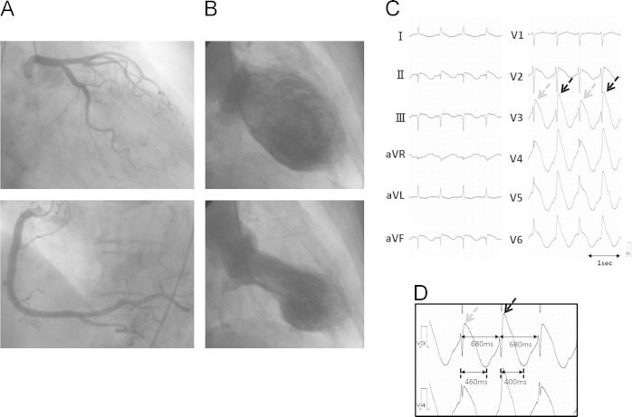
(A) Coronary angiogram showing no significant stenosis in the major coronary artery. (B) Left ventriculography revealing akinesis of the apical left ventricle and hyperkinesis of the basal left ventricle. (C and D) ECG recorded in the catheterization room. Despite stable RR intervals (680 ms), prominent J-wave and T-wave alternans were observed. The QT interval could not be measured precisely due to overlap of the terminal T wave with the following QRS. (D) shows the ECG records in V3 and V4 with an enlarged scale. The beats with higher J-wave amplitude (black arrows) had a shorter QT peak interval (400 ms) than (460 ms) beats with lower J-wave elevation (gray arrows), resulting in longer preceding diastolic interval in the latter.
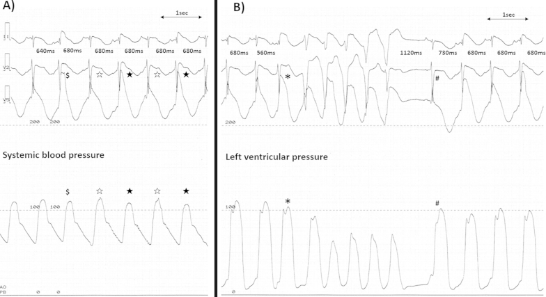
(A) The polygraphs showing simultaneous ECG recordings (II, V1, and V5) and aortic pressure. Despite a constant RR interval (680 ms), the fourth and sixth beats (black stars) had higher J-wave amplitude and lower systolic blood pressure than the third and fifth beats (white stars). The second beat ($), which had a shorter coupling interval (640 ms) had higher J-point elevation than the fourth and sixth beats (black stars). (B): The third beat (⁎), which occurred at a short coupling interval (560 ms), had prominent J-point elevation and was followed by non-sustained VT. The ninth beat (#) developed after a long coupling interval (1120 ms) and showed no J-point elevation, but induced a high ventricular pressure followed by electrical and mechanical alternans.

Serial changes in the patient׳s ECG (V1–V6) after admission. Prominent TWA and J-wave alternans were apparent until day 3 at 18:00. These changes subsequently subsided.
References
-
- Tsuchihashi K., Ueshima K., Uchida T. Angina pectoris-myocardial infarction investigations in Japan: transient left ventricular apical ballooning without coronary artery stenosis: a novel heart syndrome mimicking acute myocardial infarction. J Am Coll Cardiol. 2001;38:11–18. - PubMed
-
- Wittstein I.S., Thiemann D.R., Lima J.A. Neurohumoral features of myocardial stunning due to sudden emotional stress. N Engl J Med. 2005;352:539–548. - PubMed
-
- Samuelov-Kinori L., Kinori M., Kogan Y. Takotsubo cardiomyopathy and QT interval prolongation: who are the patients at risk for torsades de pointes? J Electrocardiol. 2009;42:353-7.e1. - PubMed
-
- Perazzolo Marra M., Zorzi A., Corbetti F. Apicobasal gradient of left ventricular myocardial edema underlies transient T-wave inversion and QT interval prolongation (Wellens׳ ECG pattern) in takotsubo cardiomyopathy. Heart Rhythm. 2013;10:70–77. - PubMed
LinkOut - more resources
Full Text Sources
Other Literature Sources
