. 2016 Sep;53(7):4821-32.
doi: 10.1007/s12035-015-9407-8. Epub 2015 Sep 4.

Examining the Neural and Astroglial Protective Effects of Cellular Prion Protein Expression and Cell Death Protease Inhibition in Mouse Cerebrocortical Mixed Cultures

Affiliations

Examining the Neural and Astroglial Protective Effects of Cellular Prion Protein Expression and Cell Death Protease Inhibition in Mouse Cerebrocortical Mixed Cultures

Kevin K W Wang et al. Mol Neurobiol. 2016 Sep.

Abstract

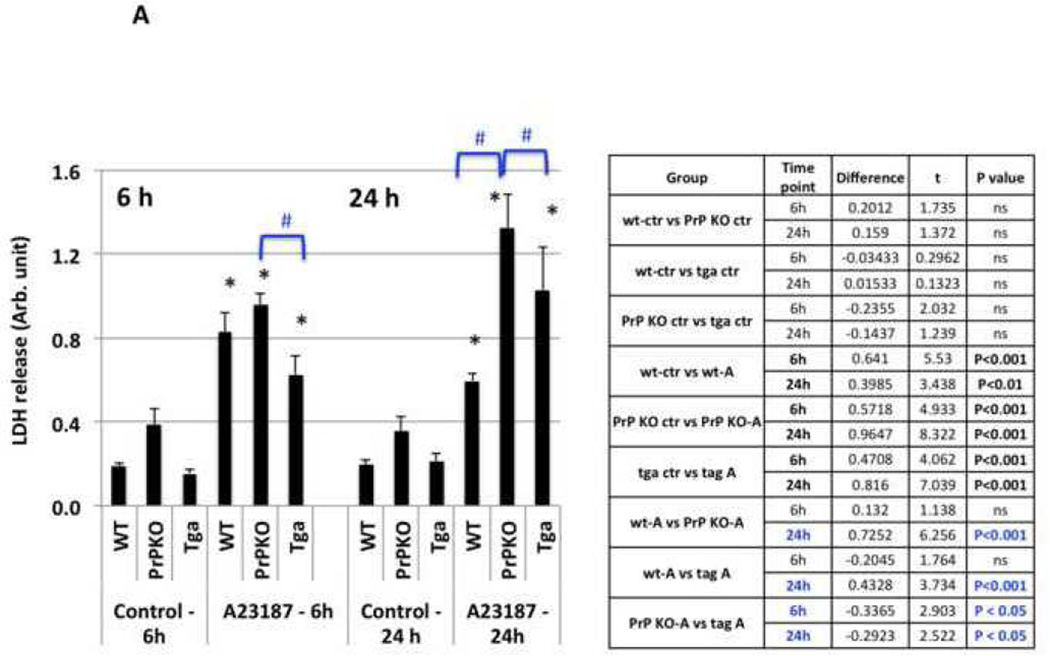
Overexpression of cellular prion protein, PrP(C), has cytoprotective effects against neuronal injuries. Inhibition of cell death-associated proteases such as necrosis-linked calpain and apoptosis-linked caspase are also neuroprotective. Here, we systematically studied how PrP(C) expression levels and cell death protease inhibition affect cytotoxic challenges to both neuronal and glial cells in mouse cerebrocortical mixed cultures (CCM). Primary CCM derived from three mouse lines expressing no (PrP(C) knockout mice (PrPKO)), normal (wild-type (wt)), or high (tga20) levels of PrP(C) were subjected to necrotic challenge (calcium ionophore A23187) and apoptotic challenge (staurosporine (STS)). CCM which originated from tga20 mice provided the most robust neuron-astroglia protective effects against necrotic and early apoptotic cell death (lactate dehydrogenase (LDH) release) at 6 h but subsequently lost its cytoprotective effects. In contrast, PrPKO-derived cultures displayed elevated A23187- and STS-induced cell death at 24 h. Calpain inhibitor SNJ-1945 protected against A23187 challenge at 6 h in CCM from all three mouse lines but protected only against A23187 and STS treatments by 24 h in the PrPKO line. In parallel, caspase inhibitor Z-D-DCB protected against pro-apoptotic STS challenge at 6 and 24 h. Furthermore, we also examined αII-spectrin breakdown products (primarily from neurons) and glial fibrillary acidic protein (GFAP) breakdown products (from astroglia) as cytoskeletal proteolytic biomarkers. Overall, it appeared that both neurons and astroglial cells were less vulnerable to proteolytic attack during A23187 and STS challenges in tga20-derived cultures but more vulnerable in PrPKO-derived cultures. In addition, calpain and caspase inhibitors provide further protection against respective protease attacks on these neuronal and glial cytoskeletal proteins in CCM regardless of mouse-line origin. Lastly, some synergistic cytoprotective effects between PrP(C) expression and addition of cell death-linked protease inhibitors were also observed.

Keywords: Apoptosis; Biomarkers; Calpain; Caspase; Cellular prion protein; Cytoprotection; Cytotoxin; Necrosis; Proteases.


Figures

Figure 1
Figure 1. Effect of PrPC expression levels on LDH release from CCM after 6 and 24 hr of cytotoxin challenge
LDH release as a monitor for cell death in wt, PrPKO and tga20 (tga) mouse-derived CCM subjected to (A) pro-necrotic challenge (20 μM A23187) and (B) pro-apoptotic challenge (1 μM staurosporine; STS) for 6hr and 24 h. Wt, PrPKO and tga20-derived CCM that are unchallenged (Control) are also included., (*) indicates LDH release levels higher than respective control. (#) indicates that PrPKO LDH release are higher or tga20 LDH levels are lower than their wt counterparts with either A23187 or STS challenge. N= 4 separate experiments. Significance is defined as p < 0.05 (two- way ANOVA). ns = not significant. Those Group comparisons that were significant are highlighted in bold. Group comparisons between two mouse lines that are significant are indicated in blue labels and #.
Figure 2
Figure 2. Effects of calpain and caspase inhibition on LDH release from CCM after 6 and 24 hr of cytotoxin challenge
LDH release as a monitor for cell death in wt, PrPKO and tga20 mouse-derived CCM subjected to pro-necrotic and pro-apoptotic challenges for 6 and 24 hr. wt (WT), PrPKO and tga20 (tga)-derived CCM are subjected to (A) pro-necrosis 20 μM A23187 [A23], (B) pro-apoptosis staurosporine [STS] with or without 30 μM SNJ-1945 (SNJ) or caspase inhibitor Z-D-DC (ZD, 50 μM). (*) indicates that calpain inhibitor SNJ-1945 (30 μM SNJ) or caspase inhibitor Z-D-DC (50 μM ZD) treatment yield statistically significant differences in LDH levels compared to the no inhibitor counterparts (with A23 or STS). N= 4 separate experiments. Significance is defined as p < 0.05 (two way ANOVA). ns = not significant. Those Group comparisons that were significant are highlighted in bold letters in the table and with * on graph.
Figure 3
Figure 3. Effect of PrPC expression on cytotoxin-induced, and neuronal injury-linked, αII-spectrin breakdown products formation in mouse CCM
Necrosis inducer A23187 (20 μM) and apoptosis inducer staurosporine (STS 1 μM) were used. (A) Representative immunoblots for αII-spectrin; quantification of (B) SBDP145 levels and (C) SBDP120 levels. Significance is defined as p < 0.05 (two-way ANOVA). ns = not significant. N= 4 separate experiments. Those Group comparisons that were significant are highlighted in bold letters in the table and with * on graph.
Figure 4
Figure 4. Effect of PrPC expression on cytotoxin-induced and glial injury-linked GFAP-BDP formation in mouse CCM
Necrosis inducer A23187 (20 μM) and apoptosis inducer staurosporine (STS 1 μM) were used. (A) Representative immunoblots for GFAP, and quantification of (B) GBDP-38K levels and (C) GBDP-22K levels. N= 4 separate experiments. Significance is defined as p < 0.05 (two-way ANOVA). ns = not significant. Those Group comparisons that were significant are highlighted in bold letters in the table and with * on graph.
Figure 5
Figure 5. Effect of calpain and caspase inhibitors on cytotoxin-induced SBDP formation in CCM from wt, PrPKO and tga20 mice
Calpain produced SBDP150, SBDP145 and caspase-produced SBDP120 were monitored. Calpain inhibitor SNJ-1945 (SNJ), caspase inhibitor Z-D-DCB (ZD) were used 1 hr prior to cytotoxin A23187 treatment for 24 h with or without SNJ or ZD (A) , or STS treatment for 24 h with or without SNJ or ZD (B). N= 4 separate experiments. Significance is defined as p < 0.05 (two- way ANOVA). ns = not significant. Those Group comparisons (A23 or STS groups with or without SNJ or ZD) that were significant are highlighted in bold. Group comparisons between two mouse lines that are significant are indicated in blue labels and #.
Figure 6
Figure 6. Effect of calpain and caspase inhibitors on cytotoxin-induced glial GFAP-BDP formation in CCM from wt, PrPKO and tga20 mice
Calpain produced GBDP-38K and putative caspase-produced GBDP-22K were monitored. (A, C) A23187 treatment for 24 h with or without SNJ-1945 or Z-D-DCB. (B, D) STS treatment for 24 h with or without SNJ-1945 or Z-D-DCB. ). N= 4 separate experiments. Significance is defined as p < 0.05 (two- way ANOVA). ns = not significant. Those Group comparisons (A23 or STS groups with or without SNJ or ZD) that were significant are highlighted in bold. Group comparisons between two mouse lines that are significant are indicated in blue labels and #.
Figure 7
Figure 7. Effects of different levels of cellular PrPc on CCM responses to A23187 and STS with or without calpain/caspase inhibition
For alphaII-spectrin, calpain-produced SBDP145 and caspase-produced SBDP120 were monitored (A, B). For GFAAP, calpain produced GBDP-38K and putative caspase-produced GBDP-22K were monitored. (C, D). N= 4 separate experiments. Significance is defined as p < 0.05 (two- way ANOVA). ns = not significant. Group comparisons between two mouse lines that are significant are highlighted in bold.

Similar articles

References

    1. Prusiner SB. The prion diseases. Brain Pathol. 1998;8:499–513. - PMC - PubMed
    1. Salès N, Rodolfo K, Hässig R, et al. Cellular prion protein localization in rodent and primate brain. Eur J Neurosci. 1998;10:2464–2471. - PubMed
    1. Caughey B, Chesebro B. Transmissible spongiform encephalopathies and prion protein interconversions. Adv Virus Res. 2001;56:277–311. - PubMed
    1. White AR, Enever P, Tayebi M, et al. Monoclonal antibodies inhibit prion replication and delay the development of prion disease. Nature. 2003;422:80–83. - PubMed
    1. Krebs B, Wiebelitz A, Balitzki-Korte B, et al. Cellular prion protein modulates the intracellular calcium response to hydrogen peroxide. J Neurochem. 2007;100:358–367. - PubMed

Publication types

MeSH terms

LinkOut - more resources