. 2015 Nov 1;26(21):3777-87.
doi: 10.1091/mbc.E15-02-0115. Epub 2015 Sep 2.

Global analysis of asymmetric RNA enrichment in oocytes reveals low conservation between closely related Xenopus species

Affiliations

Global analysis of asymmetric RNA enrichment in oocytes reveals low conservation between closely related Xenopus species

Maike Claußen et al. Mol Biol Cell. .

Abstract

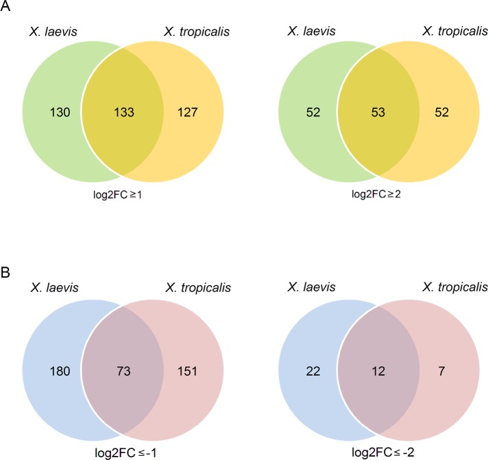
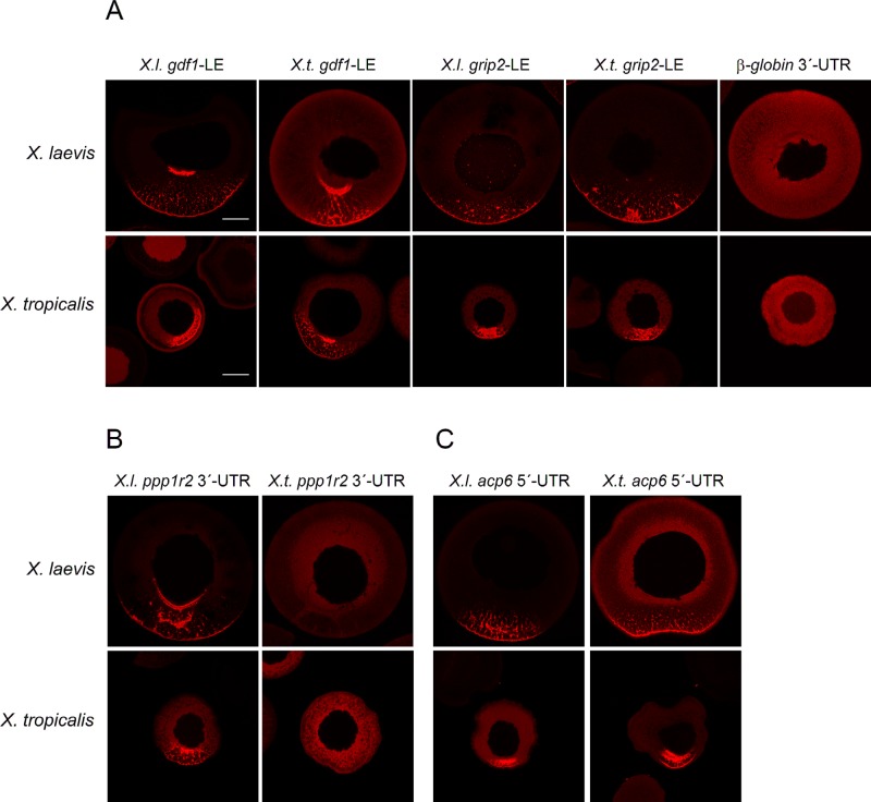
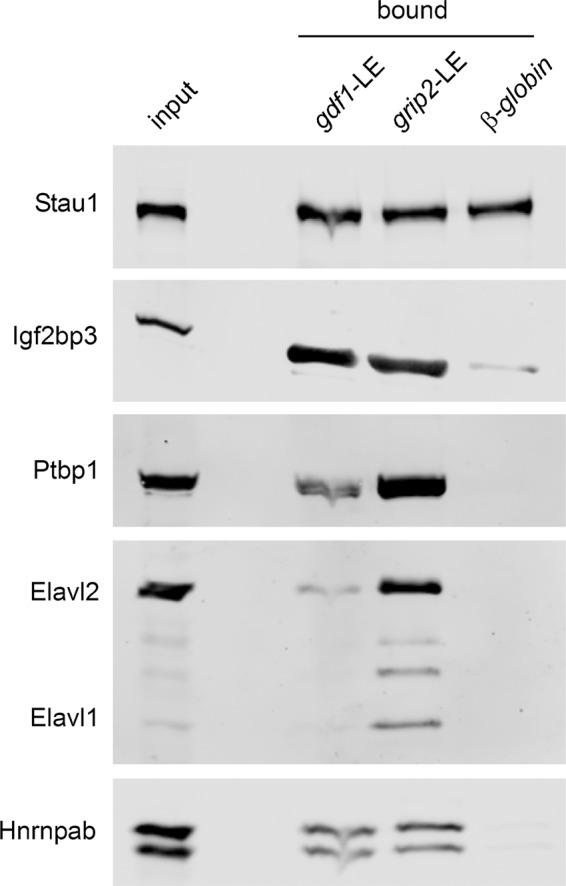
RNAs that localize to the vegetal cortex during Xenopus laevis oogenesis have been reported to function in germ layer patterning, axis determination, and development of the primordial germ cells. Here we report on the genome-wide, comparative analysis of differentially localizing RNAs in Xenopus laevis and Xenopus tropicalis oocytes, revealing a surprisingly weak degree of conservation in respect to the identity of animally as well as vegetally enriched transcripts in these closely related species. Heterologous RNA injections and protein binding studies indicate that the different RNA localization patterns in these two species are due to gain/loss of cis-acting localization signals rather than to differences in the RNA-localizing machinery.


Figures

FIGURE 1:
FIGURE 1:
Identification of novel vegetally localizing RNAs in X. laevis oocytes. (A) Candidate RNAs were tested for vegetal localization by in situ hybridization with X. laevis oocytes and listed according to their localization pattern (early and late). JgiID and gene symbol/GenBank accession number, as well as relative enrichment in the vegetal hemisphere as revealed by deep sequencing analysis (expressed as log2FC). RNAs for which no vegetal localization was detectable by in situ hybridization, as well as RNAs with very low expression levels that did not allow for the determination of localization patterns, are also listed. (B–D) Early-pathway localization pattern with characteristic mitochondrial cloud staining and spatially restricted localization at the vegetal pole (red arrows) for fnd3ca, tuft, and armc5 in stage I/II oocytes. (E–G) Late-pathway localization with typical broader vegetal cortex staining (black arrows) for slc12a9, sox7, and magi1 in stage III/IV oocytes.
FIGURE 2:
FIGURE 2:
Identification of animally enriched RNAs in X. laevis oocytes. (A) Fourteen candidate transcripts with at least fourfold animal enrichment were selected for in situ hybridization analysis with X. laevis oocytes. JgiID, gene symbol, and relative enrichment in the animal hemisphere (expressed as log2FC) as revealed by deep sequencing and qPCR analysis. Detection of animal localization by in situ hybridization is indicated. For some of the transcripts, the localization pattern could not be determined due to very low expression levels. (B–E) Animal enrichment as revealed by in situ hybridization for aen, lima, frmd8, and scl18a1 transcripts. Bisected X. laevis stage VI oocytes. Animal (a) and vegetal (v) poles.
FIGURE 3:
FIGURE 3:
Differential RNA distribution is only weakly conserved in a comparison between X. laevis and X. tropicalis. (A) Numbers of vegetally enriched RNAs from X. laevis (green) and X. tropicalis (orange) as identified by deep sequencing analysis in the form of Venn diagrams. Thresholds for vegetal localization were set to either log2FC ≥ 1 (left) or ≥ 2 (right). (B) Numbers of RNAs with animal enrichment in X. laevis (blue) and X. tropicalis (pink) oocytes in the form of Venn diagrams with thresholds set to log2FC ≤ 1 (left) and ≤ 2 (right). This comparative analysis was restricted to transcripts with expression in oocytes from both species.
FIGURE 4:
FIGURE 4:
Comparative in situ hybridization analysis confirms species-specific localization in X. laevis and X. tropicalis oocytes. (A–C) In situ hybridization with species-specific antisense RNA probes was performed with stage I–IV oocytes from X. laevis and X. tropicalis. (A) Gdf1, grip2, gplt, and cnksr2 localize to the vegetal cortex in both X. laevis and X. tropicalis oocytes. (B) Ppp1r2, pgam1, atrx, and tob2 vegetally localize in X. laevis only. (C) Mogat1, pld2, acp6, and krt8 transcripts localize to the vegetal cortex in X. tropicalis but not X. laevis oocytes.
FIGURE 5:
FIGURE 5:
Differential localization behavior of orthologous RNAs appears to rely on the RNA signal sequence but not on differences in the RNA localization machinery. (A–C) Isolated localization elements, as well as 5′-UTR, ORF, and 3′-UTRs from different transcripts and species as indicated, were labeled with cyanine-3 and injected into X. laevis and X. tropicalis oocytes. Representative confocal images of fixed oocytes. Average vegetal/animal ratios of injected RNA are listed in Supplemental Table S9. Vegetal poles are oriented toward the bottom (if assignable). Scale bars, 100 μm. (A) Injection of gdf1 and grip2 localization elements from X. laevis and X. tropicalis, as well as nonlocalizing β-globin 3′-UTR (negative control), into oocytes from both species. (B) Injection of X. laevis and X. tropicalis ppp1r2 3′-UTRs. (C) Injection of X. laevis and X. tropicalis acp6-5′-UTRs.
FIGURE 6:
FIGURE 6:
Interaction of localization proteins with LE-RNAs is conserved in X. tropicalis. Assembly of localization complexes with X. tropicalis oocyte extracts and tagged X. laevis gdf1-LE, grip2-LE, and β-globin-3′-UTR control RNA was performed in vitro. Copurifying localization proteins were detected by Western blot analysis.

References

    1. Allison R, Czaplinski K, Git A, Adegbenro E, Stennard F, Houliston E, Standart N. Two distinct Staufen isoforms in Xenopus are vegetally localized during oogenesis. RNA. 2004;10:1751–1763. - PMC - PubMed
    1. Altschul SF, Gish W, Miller W, Myers EW, Lipman DJ. Basic local alignment search tool. J Mol Biol. 1990;215:403–410. - PubMed
    1. Arthur PK, Claussen M, Koch S, Tarbashevich K, Jahn O, Pieler T. Participation of Xenopus Elr-type proteins in vegetal mRNA localization during oogenesis. J Biol Chem. 2009;284:19982–19992. - PMC - PubMed
    1. Bally-Cuif L, Schatz WJ, Ho RK. Characterization of the zebrafish Orb/CPEB-related RNA binding protein and localization of maternal components in the zebrafish oocyte. Mech Dev. 1998;77:31–47. - PubMed
    1. Bauermeister D, Claussen M, Pieler T. A novel role for Celf1 in vegetal RNA localization during Xenopus oogenesis. Dev Biol. 2015;405:214–224. - PubMed