

Correlation of magnetic resonance imaging with digital histopathology in prostate
- PMID: 26337442
- PMCID: PMC6663488
- DOI: 10.1007/s11548-015-1287-x
Correlation of magnetic resonance imaging with digital histopathology in prostate
Abstract
Purpose: We propose a systematic approach to correlate MRI and digital histopathology in prostate.
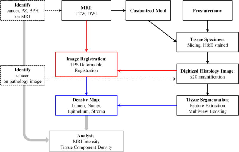
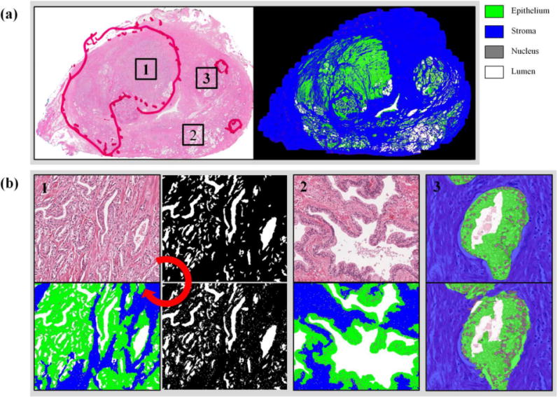
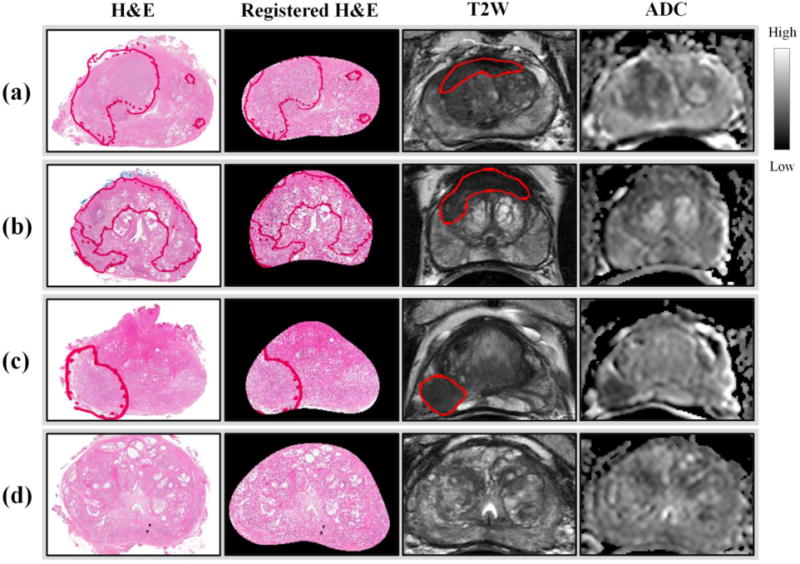
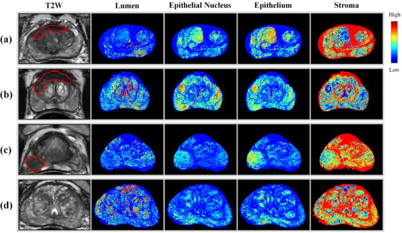
Methods: T2-weighted (T2W) MRI and diffusion-weighted imaging (DWI) are acquired, and a patient-specific mold (PSM) is designed from the MRI. Following prostatectomy, a whole mount tissue specimen is placed in the PSM and sectioned, ensuring that tissue blocks roughly correspond to MRI slices. Rigid body and thin plate spline deformable registration attempt to correct deformation during image acquisition and tissue preparation and achieve a more complete one-to-one correspondence between MRIs and tissue sections. Each tissue section is stained with hematoxylin and eosin and segmented by adopting a machine learning approach. Utilizing this tissue segmentation and image registration, the density of cellular and tissue components (lumen, nucleus, epithelium, and stroma) is estimated per MR voxel, generating density maps for the whole prostate.
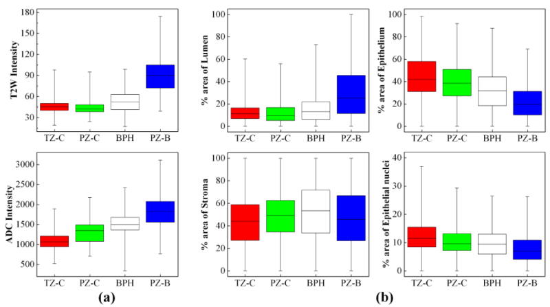
Results: This study was approved by the local IRB, and informed consent was obtained from all patients. Registration of tissue specimens and MRIs was aided by the PSM and subsequent image registration. Tissue segmentation was performed using a machine learning approach, achieving ≥0.98 AUCs for lumen, nucleus, epithelium, and stroma. Examining the density map of tissue components, significant differences were observed between cancer, benign peripheral zone, and benign prostatic hyperplasia (p value <5e−2). Similarly, the signal intensity of the corresponding areas in both T2W MRI and DWI was significantly different (p value <1e−10).
Conclusions: The proposed approach is able to correlate MRI and digital histopathology of the prostate and is promising as a potential tool to facilitate a more cellular and zonal tissue-based analysis of prostate MRI, based upon a correlative histopathology perspective.
Keywords: Histopathology; Image registration; Machine learning; Prostate.
Conflict of interest statement
Peter L. Choyke, Peter A. Pinto, and Bradford J. Wood have a cooperative research and development agreement with Philips Healthcare. Jin Tae Kwak, Sandeep Sankineni, Baris Turkbey, Sheng Xu, and Maria Merino declare that they have no conflict of interest.
Figures
References
-
- Siegel R, Ma JM, Zou ZH, Jemal A. Cancer Statistics, 2014. Ca-Cancer J Clin. 2014;64(1):9–29. - PubMed
-
- Allsbrook WC, Mangold KA, Johnson MH, Lane RB, Lane CG, Amin MB, Bostwick DG, Humphrey PA, Jones EC, Reuter VE, Sakr W, Sesterhenn IA, Troncoso P, Wheeler TM, Epstein JI. Interobserver reproducibility of Gleason grading of prostatic carcinoma: Urologic pathologists. Hum Pathol. 2001;32(1):74–80. doi: 10.1053/hupa.2001.21134. - DOI - PubMed
-
- Turkbey B, Mani H, Shah V, Rastinehad AR, Bernardo M, Pohida T, Pang YX, Daar D, Benjamin C, McKinney YL, Trivedi H, Chua C, Bratslavsky G, Shih JH, Linehan WM, Merino MJ, Choyke PL, Pinto PA. Multiparametric 3T Prostate Magnetic Resonance Imaging to Detect Cancer: Histopathological Correlation Using Prostatectomy Specimens Processed in Customized Magnetic Resonance Imaging Based Molds. J Urology. 2011;186(5):1818–1824. - PMC - PubMed
-
- Habchi H, Bratan F, Paye A, Pagnoux G, Sanzalone T, Mege-Lechevallier F, Crouzet S, Colombel M, Rabilloud M, Rouviere O. Value of prostate multiparametric magnetic resonance imaging for predicting biopsy results in first or repeat biopsy. Clin Radiol. 2014;69(3):e120–128. - PubMed
MeSH terms
Grants and funding
LinkOut - more resources
Full Text Sources
Other Literature Sources
Medical
Miscellaneous
