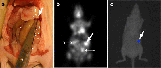
Non-invasive imaging of implanted peritoneal carcinomatosis in mice using PET and bioluminescence imaging
- PMID: 26337805
- PMCID: PMC4559549
- DOI: 10.1186/s13550-015-0125-z
Non-invasive imaging of implanted peritoneal carcinomatosis in mice using PET and bioluminescence imaging
Abstract
Background: Non-invasive imaging of peritoneal carcinomatosis remains challenging. The aim of this study was to compare positron emission tomography (PET) and bioluminescence imaging (BLI) for the early detection of peritoneal carcinomatosis in a mouse model.
Methods: Female nude mice were inoculated intraperitoneally with 1×10(7) HSC45-M2-luc gastric cancer cells. The cells were stably transfected with the gene coding for firefly luciferase. Tumour development was monitored using PET and BLI and in two subgroups, on days 3 and 4 or on days 6 and 7 after tumour cell inoculation. Tumour nodules found on post mortem examination served as the reference standard for evaluating the images.
Results: PET detected 58/82 lesions (sensitivity 71 %). This method detected all (100 %) nodules larger than 6 mm, 88 % of nodules in the range of >2-4 mm, and even 58 % of small nodules measuring only 1-2 mm. BLI identified a total of 40/82 lesions (sensitivity 49 %). The difference between PET and BLI was statistically significant at p < 0.05 (PET/BLI chi-square 8.2).
Conclusions: PET was more sensitive than BLI for the detection of early peritoneal carcinomatosis in our mouse model. The sensitivity of BLI largely depended on the site of the lesions in relation to the imaging device.
Figures


References
-
- Lordick F, Siewert JR. Recent advances in multimodal treatment for gastric cancer: a review. Gastric Cancer Off J Int Gastric Cancer Assoc Japanese Gastric Cancer Assoc. 2005;8(2):78–85. - PubMed
-
- Sadeghi B, Arvieux C, Glehen O, Beaujard AC, Rivoire M, Baulieux J, et al. Peritoneal carcinomatosis from non-gynecologic malignancies: results of the EVOCAPE 1 multicentric prospective study. Cancer. 2000;88(2):358–63. doi: 10.1002/(SICI)1097-0142(20000115)88:2<358::AID-CNCR16>3.0.CO;2-O. - DOI - PubMed
LinkOut - more resources
Full Text Sources
Other Literature Sources

