Review
. 2015;6(5):373-81.
doi: 10.1080/19491034.2015.1090073.

Moving and positioning the nucleus in skeletal muscle - one step at a time

Affiliations
Review

Moving and positioning the nucleus in skeletal muscle - one step at a time

Bruno Cadot et al. Nucleus. 2015.

Abstract

Nuclear movement and positioning within cells has become an area of great interest in the past few years due to the identification of different molecular mechanisms and functions in distinct organisms and contexts. One extreme example occurs during skeletal muscle development and regeneration. Skeletal muscles are composed of individual multinucleated myofibers with nuclei positioned at their periphery. Myofibers are formed by fusion of mononucleated myoblasts and during their development, successive nuclear movements and positioning events have been described. The position of the nuclei in myofibers is important for muscle function. Interestingly, during muscle regeneration and in some muscular diseases, nuclei are positioned in the center of the myofiber. In this review, we discuss the multiple mechanisms of nuclear positioning that occur during myofiber formation and regeneration. We also discuss the role of nuclear positioning for skeletal muscle function.

Keywords: Nuclear movement; cytoskeleton; nuclear envelope; skeletal muscle.


Figures

Figure 1.
Figure 1.
The nuclear movement multiplicity. Representative drawings of nuclear movements occurring in several cell types and organisms. Nuclei are depicted in shaded purple. In yeast, nucleus has to migrate toward the cleavage site to allow proper division. In starved fibroblasts, nuclei are positioned at the middle of the cells and move backward upon serum exposure before cell migration can proceed. After fertilization of an egg, the 2 pro-nuclei have to come in close vicinity to mix their DNAs and trigger cell division. In the vertebrate neuro-epithelium, nuclear movement is associated with cell cycle; In G1, nuclei move toward the basal side, and toward the apical side in G2. Mitosis occurs apically. During the formation of the hypodermis in C. elegans, hyp7 precursors cells has to positioned their nuclei in a face-to-face manner. Nuclear movement occurs during drosophila oogenesis, between stages6 and 8, from the posterior to the anterior edge of the oocyte. Finally, light can induce nuclear movement in leaf cells.
Figure 2.
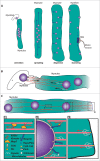
Figure 2.
Nuclear movements during muscle formation. (A) After fusion of a myoblast with a myotube, the new nucleus migrates toward the center of the myotube (centration). Then myotube nuclei get dispatched along the longest axis (spreading), followed by a migration toward the cell periphery where they become anchored (dispersion). Finally, few nuclei aggregate under the neuromuscular junction (clustering). (B) The nuclear movement after fusion involves MTs emanating from nuclei. They can bind NE through Dynein anchored by Par6. Dynein, through its ability to walk toward the minus end of MTs, will bring nuclei together. (C) The spreading movement requires MTs and can be achieved by 3 different mechanisms: 1, Anti-parallel MTs, bound to the NE through their minus end, are cross-linked by an evolutionary bridge made by the complex Map7-Kinesin-1. Kinesin-1, by walking toward the plus end of MTs will push apart nuclei. 2, Kinesin Heavy Chain bound to the nuclear envelope through Kinesin Light Chain and Nesprin can generate nuclear movements and rotations. In the same way, Dynein, bound to the nuclear envelope through Nesprin, Clip190 or Dynactin, can also generate nuclear movements. 3, Microtubules and Dynein, cortically-anchored at myotube poles through Clip190 and Raps/Pins respectively, can produce forces on plus end of microtubules. These forces will be transmitted to the NE where microtubules minus ends are anchored and then transformed in nuclear movements.

References

    1. Osorio DS, Gomes ER. The contemporary nucleus: A trip down memory lane. Biol Cell 2013; 105:430-41; PMID:23802772; http://dx.doi.org/10.1111/boc.201300009 - DOI - PubMed
    1. Sauer F. Mitosis in the neural tube. J Comp Neurol 1935; 62:377-405; http://dx.doi.org/10.1002/cne.900620207 - DOI
    1. Gundersen GG, Worman HJ. Nuclear Positioning. Cell 2013; 152:1376-89; PMID:23498944; http://dx.doi.org/10.1016/j.cell.2013.02.031 - DOI - PMC - PubMed
    1. Wilson EB. The cell in development and heredity [Internet]. MacMillan Publishing Co. 1928. [cited 2013 Aug 21] Available from: http://wellcomelibrary.org/player/b18022832
    1. Lee W-L OJ. The role of the lissencephaly protein Pac1 during nuclear migration in budding yeast. J Cell Biol 2003; 160:355-64; PMID:12566428; http://dx.doi.org/10.1083/jcb.200209022 - DOI - PMC - PubMed

Publication types

LinkOut - more resources