. 2015 Sep 2;35(35):12198-206.
doi: 10.1523/JNEUROSCI.1419-15.2015.

A Disease Mutation Causing Episodic Ataxia Type I in the S1 Links Directly to the Voltage Sensor and the Selectivity Filter in Kv Channels

Affiliations

A Disease Mutation Causing Episodic Ataxia Type I in the S1 Links Directly to the Voltage Sensor and the Selectivity Filter in Kv Channels

Dimitri Petitjean et al. J Neurosci. .

Abstract

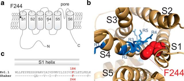
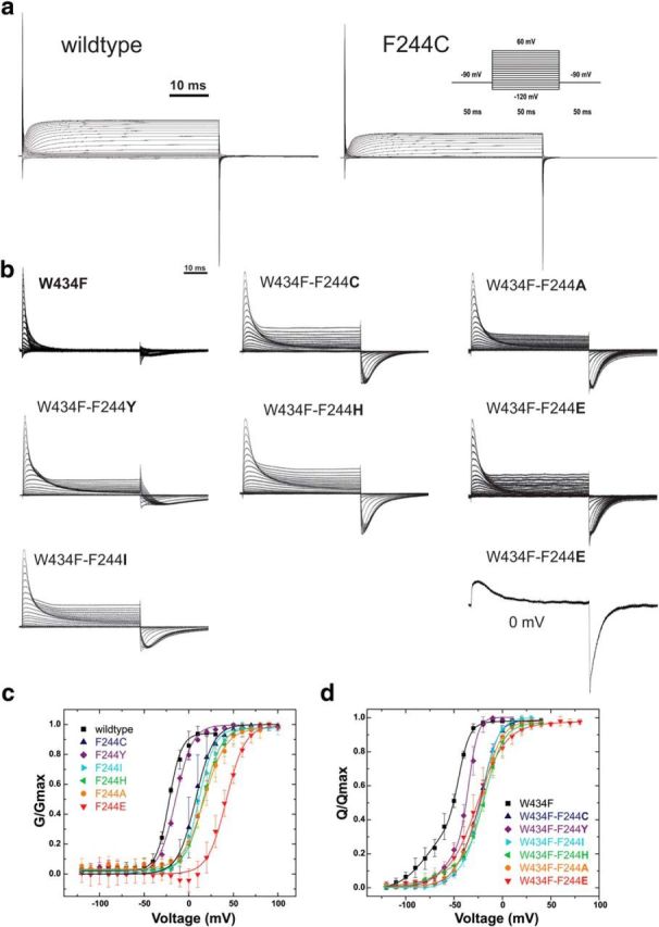
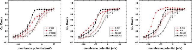
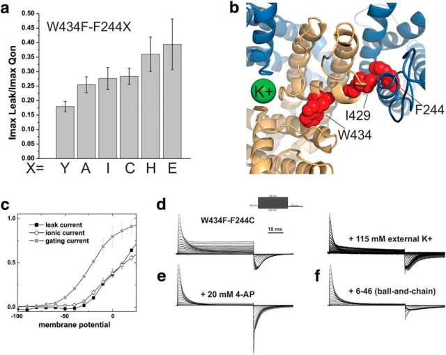
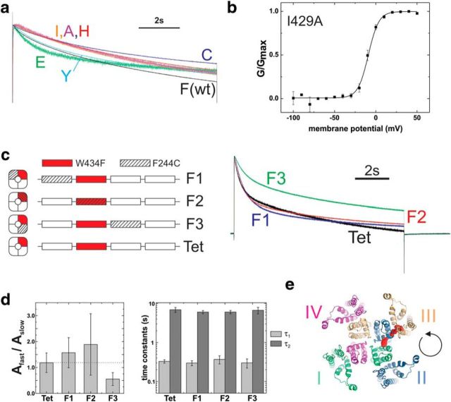
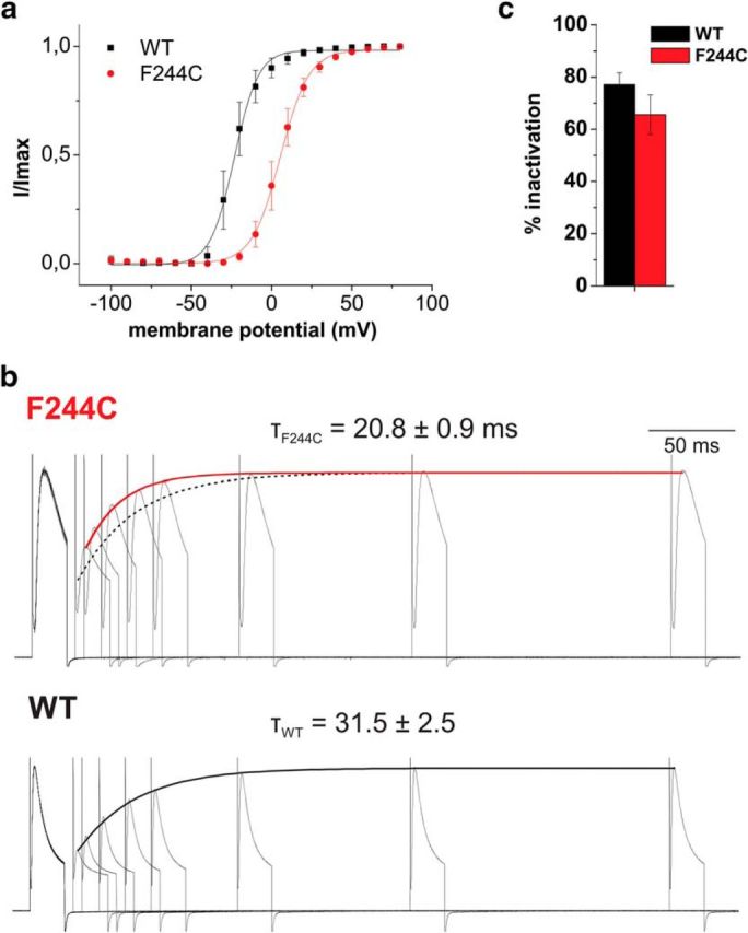
The mutation F184C in Kv1.1 leads to development of episodic ataxia type I (EA1). Although the mutation has been said to alter activation kinetics and to lower expression, we show here that the underlying molecular mechanisms may be more complex. Although F184 is positioned in the "peripheral" S1 helix, it occupies a central position in the 3D fold. We show in cut-open oocyte voltage-clamp recordings of gating and ionic currents of the Shaker Kv channel expressed in Xenopus oocytes that F184 not only interacts directly with the gating charges of the S4, but also creates a functional link to the selectivity filter of the neighboring subunit. This link leads to impaired fast and slow inactivation. The effect on fast inactivation is of an allosteric nature considering that fast inactivation is caused by a linked cytosolic ball peptide. The extensive effects of F184C provide a new mechanism underlying EA.

Significance statement: Episodic ataxia (EA) is an inherited disease that leads to occasional loss of motor control in combination with variable other symptoms such as vertigo or migraine. EA type I (EA1), studied here, is caused by mutations in a voltage-gated potassium channel that contributes to the generation of electrical signals in the brain. The mechanism by which mutations in voltage-gated potassium channels lead to EA is still unknown and there is no consistent pharmacological treatment. By studying in detail one disease-causing mutation in Kv1.1, we describe a novel molecular mechanism distinct from mechanisms described previously. This mechanism contributes to the understanding of potassium channel function in general and might lead to a better understanding of how EA develops.

Keywords: Kv channels; Xenopus oocytes; episodic ataxia; gating currents.


Figures

Figure 1.
Figure 1.
a, Topology of Kv channels F184C (F244C in Shaker) is located at the C terminus of the S1. b, Position of F244 with respect to the arginines of S4 (PDB: 3LUT). c, Sequence alignment of the S1 of Kv1.1 and Shaker Kv channels.
Figure 2.
Figure 2.
a, Current response of ShakerIR-WT and ShakerIR-F244C in response to depolarizing pulses to voltages between −120 and +60 mV from a holding potential of −90 mV. Activation is slowed in F244C. b, Gating currents in response to depolarizing pulses to voltages between −120 mV and +60 mV from a holding potential of −90 mV elicited from ShakerIR-W434F mutants and an additional mutation at position F244 as indicated. The response of F244E to a depolarizing pulse to 0 mV is shown independently. At low depolarizations, the slow onset of F244E leads to apparent larger QOFF than QON. c, GV relations of different F244 mutants elicited from protocols shown in a. d, QV relations of different F244 mutants in response to depolarizing pulses to voltages between −120 mV and +60 mV from a holding potential of −90 mV.
Figure 3.
Figure 3.
Normalized QV relations of ShakerIR-W434F (solid) and ShakerIR-F244C (open) in the absence (black) and presence (red) or neutralization of the first (R1, R362Q, left), second (R2, R365Q, center), and third (R3, R368Q, right) arginine of S4.
Figure 4.
Figure 4.
a, Ratio between maximal leak current amplitude and maximal gating current amplitude. The ratio was constant for all mutants independently of expression level. b, Position of F244 with respect to the selectivity filter. F244 of one subunit (blue) is shown in red, as well as I429 and W434 of the neighboring subunit (light orange). Potassium in the selectivity filter is shown in green (PDB: 3LUT). c, Onset of leak current (solid black squares) compared with the gating currents (solid gray squares) and the ionic current (hollow black circles). d, Gating currents of ShakerIR-W434F-F244C with K+ as the dominant ion internally and NMG (left) or K (right) as dominant ion externally. e, Block of leak currents by externally applied 4-aminopyridine. f, Block of the leak current by fast (N-type) inactivation in Shaker-W434F-F244C without deletion of positions 6–46.
Figure 5.
Figure 5.
a, Time course of slow (C-type) inactivation for ShakerIR-WT (black) and ShakerIR-F244X mutants (X = C, I, A, H, Y, E) in response to a depolarizing pulse to +60 mV from a holding potential of −90 mV. b, GV relation of ShakerIR-I429A elicited from depolarizing pulses from −90 mV to voltages between −100 mV and +50 mV. c, C-type inactivation elicited from pulses from −90 mV to +60 mV for Shaker-IR concatemers. Each concatemer contained four repeats corresponding to a Shaker-IR monomer. The W434F mutation leading to accelerated C-type inactivation is introduced in repeat II (red). Then the F244C mutation is introduced in the first (F1), second (F2), or third (F3) repeat (striped). d, Ratio of amplitudes of fast to slow component (left) and time constants (right) of exponential fits to data as shown in c. e, Illustration of the synthesis direction; based on our data, the tetramers are assembled counterclockwise when seen from the extracellular side.
Figure 6.
Figure 6.
a, Voltage dependence of fast inactivation for Shaker-WT and Shaker-F244C. Inactivation was determined by a test pulse to +50 mV (wild-type, WT) or +80 mV (F244C) after a series of depolarizations. b, Time course of recovery from inactivation for Shaker-F244C and Shaker-WT elicited from two 20 ms pulses from −90 mV to +50 mV and +80 mV for WT and F244C, respectively, with varying interpulse intervals. The dotted line marks the recovery from inactivation of Shaker-WT. c, Percentage of fast inactivation for Shaker-WT and Shaker-F244C.

References

    1. Adelman JP, Bond CT, Pessia M, Maylie J. Episodic ataxia results from voltage-dependent potassium channels with altered functions. Neuron. 1995;15:1449–1454. doi: 10.1016/0896-6273(95)90022-5. - DOI - PubMed
    1. Alle H, Geiger JR. Combined analog and action potential coding in hippocampal mossy fibers. Science. 2006;311:1290–1293. doi: 10.1126/science.1119055. - DOI - PubMed
    1. Batulan Z, Haddad GA, Blunck R. An intersubunit interaction between S4–S5 linker and S6 is responsible for the slow off-gating component in Shaker K+ channels. J Biol Chem. 2010;285:14005–14019. doi: 10.1074/jbc.M109.097717. - DOI - PMC - PubMed
    1. Baukrowitz T, Yellen G. Modulation of K+ current by frequency and external [K+]: a tale of two inactivation mechanisms. Neuron. 1995;15:951–960. doi: 10.1016/0896-6273(95)90185-X. - DOI - PubMed
    1. Berger TK, Isacoff EY. The pore of the voltage-gated proton channel. Neuron. 2011;72:991–1000. doi: 10.1016/j.neuron.2011.11.014. - DOI - PMC - PubMed

Publication types

Substances

Supplementary concepts

Associated data

LinkOut - more resources