. 2016 Feb;37(2):380-6.
doi: 10.3174/ajnr.A4504. Epub 2015 Sep 3.

Clinical Outcomes of Patients with Delayed Diagnosis of Spinal Dural Arteriovenous Fistulas

Affiliations

Clinical Outcomes of Patients with Delayed Diagnosis of Spinal Dural Arteriovenous Fistulas

W Brinjikji et al. AJNR Am J Neuroradiol. 2016 Feb.

Abstract

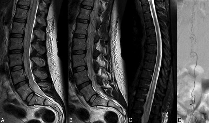
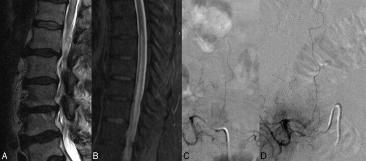
Background and purpose: Spinal dural arteriovenous fistulas are commonly missed on imaging or misdiagnosed as inflammatory or neoplastic processes. We reviewed a consecutive series of spinal dural arteriovenous fistulas referred to our institution that were missed or misdiagnosed on initial imaging and studied the clinical consequences of missing or misdiagnosing the lesion.

Materials and methods: We reviewed spinal dural arteriovenous fistulas diagnosed at our institution between January 1, 2000, and November 1, 2014. A lesion was defined as "misdiagnosed" if initial MR imaging or CT myelography demonstrated characteristic imaging features of spinal dural arteriovenous fistula but the patient was clinically or radiologically misdiagnosed. Outcomes included length of delay of diagnosis, increased disability (increase in mRS or Aminoff motor disability of ≥1 point) between initial imaging evaluation and diagnosis date, and posttreatment disability.

Results: Fifty-three consecutive spinal dural arteriovenous fistulas that were initially misdiagnosed despite having characteristic imaging findings on MR imaging or CT myelography were included in our study. Eight patients (18.9%) underwent spinal angiography before referral, which was interpreted as having negative findings but was either incomplete (6 cases) or retrospectively demonstrated the spinal dural arteriovenous fistulas (2 cases). The median time of delayed diagnosis was 6 months (interquartile range, 2-14 months). Fifty-one patients (96.2%) had increased disability between the initial study, which demonstrated features of a spinal dural arteriovenous fistula, and diagnosis. Thirty-two patients (60.4%) developed a new requirement for a walker or wheelchair. Following treatment, 21 patients (41.2%) had an improvement of 1 point on the mRS or Aminoff motor disability scale.

Conclusions: Delayed diagnosis of spinal dural arteriovenous fistula with characteristic imaging features results in high rates of additional disability that are often irreversible despite surgical or endovascular treatment of the fistula.


Figures

Fig 1.
Fig 1.
A 57-year-old woman with a 3-month history of bilateral lower extremity tingling and progressive lower extremity weakness. A and B, T2-weighted lumbar spine MR images demonstrate high T2 signal in the conus with multiple flow voids in the intradural space. C, T2-weighted MR image of the thoracic spine demonstrates high T2 signal in the lower thoracic cord to the conus. The patient was diagnosed with neuromyelitis optica and received no spinal-vasculature imaging before referral to our institution. Two rounds of IV methylprednisolone (Solu-Medrol) therapy resulted in worsening of symptoms, and rituximab therapy was of no benefit. D, Spinal angiography demonstrates the spinal dural AVF with an arterial feeder from the L3 radiculomeningeal artery.
Fig 2.
Fig 2.
A 68-year-old man with a 3-month history of saddle anesthesia, constipation, difficulty voiding, and numbness in the lower extremities. T2-weighted images of the lumbar and thoracic spine demonstrate high T2 signal in the lower thoracic cord and conus (A and B). Due to clinical suspicion of SDAVF, an angiogram was obtained before referral to our center. C, The angiogram clearly demonstrates the fistula arising from the L2 radiculomeningeal artery; however, it was interpreted as a negative finding. Before the diagnosis was made, the patient underwent an extensive imaging and clinical evaluation, including a panel negative for paraneoplastic syndrome, PET/CT, and lumbar puncture. Two rounds of IV Solu-Medrol therapy resulted in worsening of symptoms. The patient also underwent a T10–T11 laminectomy and 2 spinal cord biopsies. D, Repeat spinal angiography re-demonstrates the fistula.

References

    1. Marcus J, Schwarz J, Singh IP, et al. . Spinal dural arteriovenous fistulas: a review. Curr Atheroscler Rep 2013;15:335 10.1007/s11883-013-0335-7 - DOI - PubMed
    1. Iovtchev I, Hiller N, Ofran Y, et al. . Late diagnosis of spinal dural arteriovenous fistulas resulting in severe lower-extremity weakness: a case series. Spine J 2015;15:e39–44 10.1016/j.spinee.2013.08.029 - DOI - PubMed
    1. Morris JM. Imaging of dural arteriovenous fistula. Radiol Clin North Am 2012;50:823–39 10.1016/j.rcl.2012.04.011 - DOI - PubMed
    1. Abe T, Tokuda Y, Ishimatsu S, et al. . Spinal dural arteriovenous fistula incidentally discovered. J Emerg Trauma Shock 2011;4:299 10.4103/0974-2700.82227 - DOI - PMC - PubMed
    1. Aghakhani N, Parker F, David P, et al. . Curable cause of paraplegia: spinal dural arteriovenous fistulae. Stroke 2008;39:2756–59 10.1161/STROKEAHA.108.517037 - DOI - PubMed

LinkOut - more resources