. 2015 Nov;89(22):11438-56.
doi: 10.1128/JVI.01794-15. Epub 2015 Sep 2.

The Genome of a Tortoise Herpesvirus (Testudinid Herpesvirus 3) Has a Novel Structure and Contains a Large Region That Is Not Required for Replication In Vitro or Virulence In Vivo

Affiliations

The Genome of a Tortoise Herpesvirus (Testudinid Herpesvirus 3) Has a Novel Structure and Contains a Large Region That Is Not Required for Replication In Vitro or Virulence In Vivo

Frédéric Gandar et al. J Virol. 2015 Nov.

Abstract

Testudinid herpesvirus 3 (TeHV-3) is the causative agent of a lethal disease affecting several tortoise species. The threat that this virus poses to endangered animals is focusing efforts on characterizing its properties, in order to enable the development of prophylactic methods. We have sequenced the genomes of the two most studied TeHV-3 strains (1976 and 4295). TeHV-3 strain 1976 has a novel genome structure and is most closely related to a turtle herpesvirus, thus supporting its classification into genus Scutavirus, subfamily Alphaherpesvirinae, family Herpesviridae. The sequence of strain 1976 also revealed viral counterparts of cellular interleukin-10 and semaphorin, which have not been described previously in members of subfamily Alphaherpesvirinae. TeHV-3 strain 4295 is a mixture of three forms (m1, m2, and M), in which, in comparison to strain 1976, the genomes exhibit large, partially overlapping deletions of 12.5 to 22.4 kb. Viral subclones representing these forms were isolated by limiting dilution assays, and each replicated in cell culture comparably to strain 1976. With the goal of testing the potential of the three forms as attenuated vaccine candidates, strain 4295 was inoculated intranasally into Hermann's tortoises (Testudo hermanni). All inoculated subjects died, and PCR analyses demonstrated the ability of the m2 and M forms to spread and invade the brain. In contrast, the m1 form was detected in none of the organs tested, suggesting its potential as the basis of an attenuated vaccine candidate. Our findings represent a major step toward characterizing TeHV-3 and developing prophylactic methods against it.

Importance: Testudinid herpesvirus 3 (TeHV-3) causes a lethal disease in tortoises, several species of which are endangered. We have characterized the viral genome and used this information to take steps toward developing an attenuated vaccine. We have sequenced the genomes of two strains (1976 and 4295), compared their growth in vitro, and investigated the pathogenesis of strain 4295, which consists of three deletion mutants. The major findings are that (i) TeHV-3 has a novel genome structure, (ii) its closest relative is a turtle herpesvirus, (iii) it contains interleukin-10 and semaphorin genes (the first time these have been reported in an alphaherpesvirus), (iv) a sizeable region of the genome is not required for viral replication in vitro or virulence in vivo, and (v) one of the components of strain 4295, which has a deletion of 22.4 kb, exhibits properties indicating that it may serve as the starting point for an attenuated vaccine.


Figures

FIG 1
FIG 1
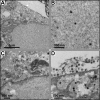
Morphology of TeHV-3. TH-1 cells were infected with strains 1976 and 4295. Six days postinfection, cells were processed for electron microscopy examination. In a blind test, it was not possible to differentiate the two strains. The images represent TH-1 cells infected with strain 1976. (A) General view of the various compartments of an infected cell; (B) nucleoplasm; (C) nucleus and cytoplasm; (D) cytoplasm and extracellular space.
FIG 2
FIG 2
Map of the TeHV-3 strain 1976 genome. The unique regions (UT, UL, and US) are in white, and the inverted repeats (TRT, IRT, IRS, and TRS) are in light yellow. Predicted functional ORFs are depicted by colored arrows, with nomenclature below them. Red shading indicates ORFs inherited from the ancestor of the alpha-, beta-, and gammaherpesviruses. Blue shading indicates ORFs that have orthologs in mammalian or avian alphaherpesviruses. Light orange shading indicates the remaining ORFs. Introns are shown as narrow white bars. Reiterations are shown by gray shading within or between ORFs, and three copies of ori are indicated by vertical red bars. The deletions and the duplications present in the m1, m2, and M forms of TeHV-3 strain 4295 are marked above the genome by horizontal green and orange bars, respectively.
FIG 3
FIG 3
Relative orientations of unique regions in the TeHV-3 strain 1976 genome. (A) A schematic representation of the genome is shown at the top, the orientations of UT, UL, and US corresponding to those in Fig. 2. Below this, the four possible combinations of the orientations of UT-UL and UL-US are presented, with the majority of UL omitted. For each combination, the sizes (in kilobases) of restriction endonuclease fragments at or near the genome ends or the UT-UL and UL-US junctions are shown (EcoRI above the genome and KpnI below). White bars (P1 to P6) represent the positions of the probes used for hybridization. Asterisks highlight restriction endonuclease fragments for which a positive band was observed in Southern blot analyses. (B) The panels on the right show a Southern blot analysis of TeHV-3 strain 1976 DNA digested with EcoRI or KpnI and hybridized to probes P1 to P6. Black arrowheads indicate all bands compatible with the predicted fragments. The panel on the left shows an ethidium bromide-stained profile of marker fragments (MS) and strain 1976 DNA digested with EcoRI or KpnI. The 0.65- and 0.72-kb fragments were detected but are not visible on these images, which have been restricted to fragments above 2 kb.
FIG 4
FIG 4
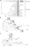
Phylogenetic analyses. In each panel, the scale indicates substitutions per site. Abbreviations for herpesvirus names: El, elephantid; H, human; Mu, murid; E, equid; F, felid; Bo, bovid; S, suid; Ce, cercopithecine; Pt, pteropodid; Pn, panine; Mc, macacine; Pa, papiine; Sa, saimiriine; Ga, gallid; Me, meleagrid; Co, columbid; An, anatid; Ac, accipitrid; Ps, psittacid; Te, testudinid; Ch, chelonid; Ov, ovine; Al, alcelaphine; Cy, cyprinid; and HV, herpesvirus (followed by a hyphenated number). Other abbreviations: CNPV, canarypox virus; FPV, fowlpox virus; PEPV, penguin poxvirus; PNPV, pigeon poxvirus; SGIV, Singapore grouper iridovirus; VARV, variola virus; HSPV, horse poxvirus; VACV, vaccinia virus; ECTV, ectromelia virus; CPXV, cowpox virus; RPXV, rabbit poxvirus; ORFV, ORF virus; and SWPV, swinepox virus. (A) Bayesian tree for herpesvirus DNA polymerase proteins. All nodes have posterior probabilities of 1. Viruses that have not yet been classified are marked by asterisks (1). (B) Maximum likelihood tree for vertebrate SEMA-7A proteins and their viral homologs. Node bootstrap values greater than 70 are marked. (C) Maximum likelihood tree for IL-10 homologs. Node bootstrap values greater than 70 are marked.
FIG 5
FIG 5
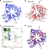
Homology model of the TeHV-3 SEMA (TE7) protein. (A) Mouse SEMA-3A PDB 1Q47. (B) Homology model of TeHV-3 TE7 constructed from PDB 1Q47 with suboptimal residues shown. (C) Ramachandran plot of homology model used to identify suboptimal residues. (D) Mouse SEMA-3A PDB 1Q47 (blue) superposed with the homology model of TeHV-3 TE7 (red).
FIG 6
FIG 6
Homology model of the TeHV-3 IL-10 (TE8) protein. (A) Human IL-10 PDB 1ILK. (B) Homology model of TeHV-3 IL-10 constructed from 1ILK with suboptimal residues shown. (C) Ramachandran plot of homology model used to identify suboptimal residues. (D) Human IL-10 PDB 1ILK (blue) superposed with the homology model of TeHV-3 IL-10 (red).
FIG 7
FIG 7
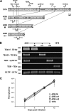
Effects of the deletions in the m1, m2, and M forms of strain 4295 on viral growth in vitro. (A) Schematic representation of the regions in the strain 1976 genome corresponding to the deletions in the three forms of strain 4295, with the coordinates of the deletions indicated above the strain 1976 genome. Arrows represent primers (Table 1) designed for PCR amplification of the regions containing the deletions. Amplicon sizes are indicated in bold below the genomes of the strain 4295 subclones. PCR amplification performed with the UL13f and UL13r primers led to a product of 106 bp. (B) Characterization by PCR of the strain 4295 subclones representing the three genome forms. Strain 4295 prior to subcloning (containing the three forms) and strain 1976 were used as controls. The positions of markers (in base pairs) are marked by arrowheads. (C) Multistep growth curves of the strain 4295 subclones and strain 1976.
FIG 8
FIG 8
Pathogenesis of strain 4295. On day 0, tortoises (n = 4) aged 5.4 years (mean weight ± SD, 219.5 g ± 53.1 g) were infected (n = 3) with strain 4295 (consisting of a mixture of three deletion mutants) or mock-infected (n = 1). Percentage survival is expressed according to the days postinfection. The tortoises were named according to the following scheme: the inoculation performed (strain 4295 or mock)/the time postinfection at which death occurred (when more than one tortoise died on the same day, they were further ranked by the addition of a lowercase letter) and an uppercase letter to describe the clinical state before euthanasia (D, diseased; H, healthy).
FIG 9
FIG 9
Histopathological characterization of the lesions induced by strain 4295 (consisting of a mixture of three deletion mutants). The indicated organs were collected from all tortoises at the end of the experiment described in the legend to Fig. 8 and were processed for histological examination. The images were collected from tortoises Mock/75 H and 4295/41 D, the latter having been selected as representative of the infected group. H, heterophil; F, faveolae; GC, glial cell; C, capillary; PT, proximal tubule; DT, distal tubule; G, glomerulus; S, sinusoid; and M, melanomacrophage. Bars, 20 μm.
FIG 10
FIG 10
Tissue tropism of strain 4295. DNA was extracted from the indicated organs of all tortoises at the end of the experiment described in the legend of Fig. 8. (A) Analysis of viral gene copy number by qPCR. Individual values represent the means from triplicate measurements ± SD. (B) Analysis of the presence of the three forms present in strain 4295, using primers specific for each form (m1, m2, and M) and all three forms (UL13). See Fig. 7A and B and Table 1 for details of the primers. Strain 4295 (containing the three forms) was used as positive (CT) control.

References

    1. Pellett PE, Davison AJ, Eberle R, Ehlers B, Hayward GS, Lacoste V, Minson AC, Nicholas J, Roizman B, Studdert MJ, Wang F. 2012. Order—Herpesvirales, p 99–107. In King AMQ, Adams MJ, Carstens EB, Lefkowitz EJ (ed), Virus taxonomy. Elsevier, San Diego, CA. doi:10.1016/B978-0-12-384684-6.00005-7. - DOI
    1. Pellett PE, Roizman B. 2013. Herpesviridae, p 1802–1822. In Knipe DM, Howley PM (ed), Fields virology, 6th ed, vol 2 Lippincott Williams & Wilkins, Philadelphia, PA.
    1. Bicknese EJ, Childress AL, Wellehan JF Jr. 2010. A novel herpesvirus of the proposed genus Chelonivirus from an asymptomatic bowsprit tortoise (Chersina angulata). J Zoo Wildl Med 41:353–358. doi:10.1638/2009-0214R.1. - DOI - PubMed
    1. Ackermann M, Koriabine M, Hartmann-Fritsch F, de Jong PJ, Lewis TD, Schetle N, Work TM, Dagenais J, Balazs GH, Leong JA. 2012. The genome of Chelonid herpesvirus 5 harbors atypical genes. PLoS One 7:e46623. doi:10.1371/journal.pone.0046623. - DOI - PMC - PubMed
    1. Origgi FC. 2006. Herpesvirus in tortoises, p 814–821. In Mader DR. (ed), Reptile medicine and surgery, 2nd ed Saunders, St. Louis, MO.

Publication types

MeSH terms

LinkOut - more resources