

A rare case of pulmonary sclerosing hemagioma with lymph node metastasis and review of the literature
- PMID: 26339444
- PMCID: PMC4555772
A rare case of pulmonary sclerosing hemagioma with lymph node metastasis and review of the literature
Abstract
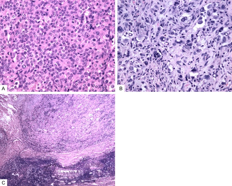
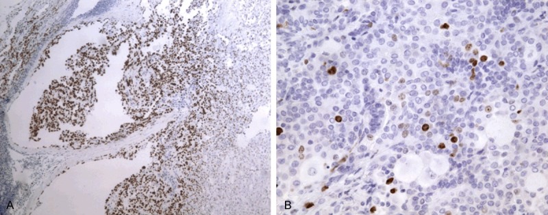
Pulmonary sclerosing hemagioma (SH) is an uncommon tumor with malignance potential. Clinically this disease is regarded as benign but extremely rare cases can have lymph node metastasis. Up to date, there have been only very few reports concerning SH with lymph node metastasis. In this paper we reported one pulmonary SH case with lymph node metastasis and additionally overviewed the clinical and pathological features of SH. A young-aged female was found incidentally to have a nodule in the right upper lung. This patient presented no cough, no hemoptysis and chest pain. Computed tomography (CT) scan indicated a large mass in the right upper lung and enlarged lymph nodes in the right hilum. The patient underwent lobectomy of the right upper lung. Histologically, the tumor demonstrated typical features of SH and was consisted of angiomatoid areas, sclerosis, papillary structures lined with cuboidal cells and sheets of round to polygonal cells. Polygonal cells in some solid areas presented abnormal enlarged nuclei and increased karyoplasmic ratio; tumor giant cells were noted; whereas mitosis was not observed. One peribronchial lymph node was noted for SH metastasis and the metastatic tissue were consisted of polygonal cells. Immunohistochemistry (IHC) revealed that both surface-lining cuboidal and polygonal cells expressed EMA and thyroid transcription factor 1 (TTF-1), but were negative for CD34, VIII factor, CD68 and Claratinin. The polygonal cells showed relatively higher expression of Ki-67 and p53 than the surface-lining cells. Postoperatively, the patient received no chemotherapy or radiotherapy and no recurrence 2 years after surgery was noted.
Keywords: Sclerosing hemangioma (SH); lymph node metastasis; pulmonary SH.
Figures
References
-
- Liebow A, Hubbell DS. Sclerosing hemangioma (histocytoma, xarthoma) of the lung. Cancer. 1956;9:53–75. - PubMed
-
- Wang Y, Wang EH, Wu GP, Zhang ZK, Lin D. Pulmonary sclerosing hemangioma immunochemical markers and ultra-structure study: different progenitors of SH. Chinese Journal of Lung Cancer. 2003;6:89–93. - PubMed
-
- Jiang ZN, Zhu T, Jin M, Wang LB. Case report: Pulmonary sclerosing hemangioma with lymph node metastasis. Chinese Journal of Pathology. 2007;36:282–283. - PubMed
-
- Chan AC, Chan JK. Pulmonary sclerosing hemangioma consistently expresses thyroid transcription factor-1 (TTF-1): a new clue to its histogenesis. Am J Surg Pathol. 2000;24:1531–1536. - PubMed
-
- Devouassoux-Shisheboran M, Hayashi T, Linnoila RI, Koss MN, Travis WD. A clinicopathologic study of 100 cases of pulmonary sclerosing hemangioma with immunohistochemical studies: TTF-1 is expressed in both round and surface cells, suggesting an origin from primitive respiratory epithelium. Am J Surg Pathol. 2000;24:906–916. - PubMed
Publication types
MeSH terms
Substances
LinkOut - more resources
Full Text Sources
Research Materials
Miscellaneous